. 2022 Jun;23(6):420.
doi: 10.3892/etm.2022.11347. Epub 2022 Apr 29.

Simvastatin alleviates epithelial-mesenchymal transition and oxidative stress of high glucose-induced lens epithelial cells in vitro by inhibiting RhoA/ROCK signaling

Affiliations

Simvastatin alleviates epithelial-mesenchymal transition and oxidative stress of high glucose-induced lens epithelial cells in vitro by inhibiting RhoA/ROCK signaling

Jianming Fu et al. Exp Ther Med. 2022 Jun.

Abstract

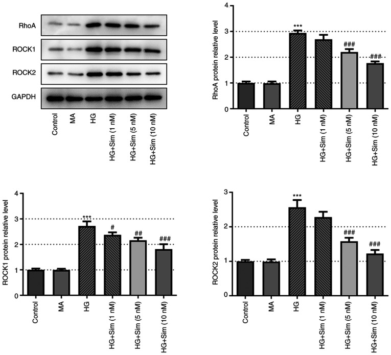
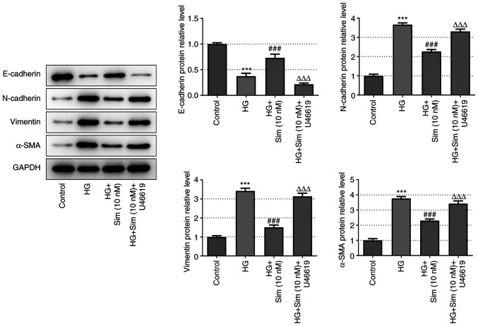
Diabetic cataracts (DC) is one of the main causes of blindness among patients with diabetes mellitus. The aim of the present study was to examine the effect of simvastatin on lens epithelial cells in DC and the underlying mechanism. The viability of SRA01/04 cells treated with different concentrations of simvastatin was detected using a Cell Counting Kit-8 assay before and after high glucose (HG) treatment. The expression levels of E-cadherin, N-cadherin, Vimentin and α-smooth muscle actin (α-SMA), proteins associated with epithelial-mesenchymal transition (EMT), in addition to RhoA, Rho-associated kinases (ROCK)1 and ROCK2, proteins related to RhoA/ROCK signaling, were also measured in SRA01/04 cells treated with HG and simvastatin, with or without U46619, using western blot analysis. DCFH-DA dyes, superoxide dismutase (SOD) and glutathione (GSH)/glutathione disulfide (GSSG) kits were used to measure the levels of oxidative stress parameters in SRA01/04 cells treated with HG and simvastatin with or without U46619. The cell viability of SRA01/04 cells treated with simvastatin was found to be significantly elevated after HG treatment. The protein expression levels of E-cadherin were increased but those of N-cadherin, Vimentin and α-SMA decreased after HG and simvastatin treatment, and this was reversed by U46619. The levels of SOD and GSH-GSSG were found to be increased whereas reactive oxygen species levels were decreased, effects that were reversed by U46619. Additionally, the protein expression levels of RhoA, ROCK1 and ROCK2 were markedly decreased. These findings provided evidence that simvastatin increased HG-induced SRA01/04 cell viability and exerted inhibitory effects on EMT and oxidative stress that occurs during DC.

Keywords: RhoA/Rho-associated protein kinase; diabetic cataract; epithelial-mesenchymal transition; oxidative stress; simvastatin.


Conflict of interest statement

The authors declare that they have no competing interests.

Figures

Figure 1
Figure 1
Simvastatin enhanced the cell viability of human lens epithelial cells SRA01/04 induced by HG. (A) The chemical formula of simvastatin. (B) The viability of SRA01/04 cells treated with different concentrations of simvastatin for 48 h was examined by CCK-8. (C) The viability of SRA01/04 cells treated with different concentrations of simvastatin for 48 h was tested by means of CCK-8 after adding different doses of HG. Data are expressed as mean ± SD. **P<0.001 vs. Control. #P<0.05 vs. HG. HG, high glucose; SD, standard deviation.
Figure 2
Figure 2
Simvastatin inhibited the EMT of SRA01/04 cells induced by HG. (A) Protein levels of E-cadherin, N-cadherin, Vimentin and α-SMA which were related to EMT were detected by western blot analysis. (B) Quantification of the related bands. Data are expressed as mean ± SD. ***P<0.001 vs. Control. #P<0.05, ##P<0.01, ###P<0.001 vs. HG. HG, high glucose; SD, standard deviation.
Figure 3
Figure 3
Simvastatin alleviated the oxidative stress of SRA01/04 cells induced by HG. (A) Level of ROS was measured by means of DCFH-DA kit after adding 25 nM HG and different doses of simvastatin. Green-stained cells are positive cells. Scale bar: 50 µm. (B) The level of SOD was detected with the corresponding SOD kit after adding 25 nM HG and different doses of simvastatin. (C) The GSH-GSSG level was examined with GSH-GSSG kit after adding 25 nM HG and different doses of simvastatin. Data are expressed as mean ± SD. ***P<0.001 vs. Control. #P<0.05, ###P<0.001 vs. HG. HG, high glucose; SD, standard deviation.
Figure 4
Figure 4
Simvastatin suppressed RhoA/ROCK signaling transduction. The protein levels of RhoA, ROCK1 and ROCK2 were measured by western blot analysis after treatment with HG and different doses of simvastatin. Data are expressed as mean ± SD. ***P<0.001 vs. Control. #P<0.05, ##P<0.01, ###P<0.001 vs. HG. HG, high glucose; SD, standard deviation.
Figure 5
Figure 5
Simvastatin inhibited the EMT of SRA01/04 cells induced by HG by suppressing RhoA/ROCK signaling transduction. Protein levels of E-cadherin, N-cadherin, Vimentin and α-SMA were detected by western blot analysis after the treatment of HG, 10 nM simvastatin and the activator of RhoA/ROCK pathway U46619. Data are expressed as mean ± SD. ***P<0.001 vs. Control. ###P<0.001 vs. HG. ΔΔΔP<0.001 vs. HG + Sim (10 nM). HG, high glucose; Sim, simvastatin; SD, standard deviation.
Figure 6
Figure 6
Simvastatin alleviated the oxidative stress of SRA01/04 cells induced by high glucose via suppressing RhoA/ROCK signaling transduction. (A) The level of ROS was measured by means of DCFH-DA kit after adding 10 nM U46619. Green-stained cells are positive cells. Scale bar: 50 µm. (B) The level of SOD was detected with the corresponding SOD kit after adding 10 nM U46619. (C) The GSH-GSSG level was examined with GSH-GSSG kit after adding 10 nM U46619. Data are expressed as mean ± SD. ***P<0.001 vs. Control. ###P<0.001 vs. HG. ΔΔP<0.01, ΔΔΔP<0.001 vs. HG + Sim (10 nM). HG, high glucose; Sim, simvastatin; SD, standard deviation.

Similar articles

Cited by

References

    1. Cole JB, Florez JC. Genetics of diabetes mellitus and diabetes complications. Nat Rev Nephrol. 2020;16:377–390. doi: 10.1038/s41581-020-0278-5. - DOI - PMC - PubMed
    1. Ye W, Ma J, Wang F, Wu T, He M, Li J, Pei R, Zhang L, Wang Y, Zhou J. LncRNA MALAT1 regulates miR-144-3p to facilitate epithelial-mesenchymal transition of lens epithelial cells via the ROS/NRF2/Notch1/snail pathway. Oxid Med Cell Longev. 2020;2020(8184314) doi: 10.1155/2020/8184314. - DOI - PMC - PubMed
    1. Sreedharan R, Abdelmalak B. Diabetes mellitus: Preoperative concerns and evaluation. Anesthesiol Clin. 2018;36:581–597. doi: 10.1016/j.anclin.2018.07.007. - DOI - PubMed
    1. Panozzo G, Staurenghi G, Dalla Mura G, Giannarelli D, Alessio G, Alongi S, Appolloni R, Baldascino A, Boscia F, Caporossi A, et al. Prevalence of diabetes and diabetic macular edema in patients undergoing senile cataract surgery in Italy: The DIabetes and CATaract study. Eur J Ophthalmol. 2020;30:315–320. doi: 10.1177/1120672119830578. - DOI - PMC - PubMed
    1. Andley UP. The lens epithelium: Focus on the expression and function of the alpha-crystallin chaperones. Int J Biochem Cell Biol. 2008;40:317–323. doi: 10.1016/j.biocel.2007.10.034. - DOI - PMC - PubMed