. 2022 May 4:12:882843.
doi: 10.3389/fcimb.2022.882843. eCollection 2022.

Nucleospora hippocampi n. sp., an Intranuclear Microsporidian Infecting the Seahorse Hippocampus erectus From China

Affiliations

Nucleospora hippocampi n. sp., an Intranuclear Microsporidian Infecting the Seahorse Hippocampus erectus From China

Yuan Wang et al. Front Cell Infect Microbiol. .

Abstract

The life cycle, ultrastructure, and molecular phylogeny of a new intranuclear microsporidian, Nucleospora hippocampi n. sp., infecting the intestine of the Hippocampus erectus, were described. The histopathology revealed an extensive infection, mainly in the columnar epithelium of the intestinal mucosa layer. The enterocytes were the important target cell for Nucleospora hippocampi n. sp. infection. Transmission electron microscopy results showed that this microsporidian developed directly within the host cell nucleoplasm. In the intranuclear life cycle, the transformation from meront to sporogonial plasmodium was recognized by forming electron-dense disc structures, which were considered the polar tube precursors. The microsporidian showed the typical morphological characteristics of the family Enterocytozoonidae in the formation and development of spore organelles prior to the division of the sporogonial plasmodium. According to wet smear observation, eight spores were generally formed in a single host nucleus. Mature spores were elongated ovoids that were slightly bent and measured 1.93 × 0.97 μm. The isofilar polar tube was arranged in 7~8 coils in one row. Phylogenetic analysis of its small subunit ribosomal DNA sequences demonstrated that the parasite belonged to the Nucleospora group clade. The histological, ultrastructural, and molecular data support the emergence of a new species in the genus Nucleospora. This is the first report of Nucleospora species in Asia and threatened syngnathid fishes.

Keywords: Nucleospora; intestinal disease; intranuclear parasitism; seahorse; transmission electron microscopy.


Conflict of interest statement

The authors declare that the research was conducted in the absence of any commercial or financial relationships that could be construed as a potential conflict of interest.

Figures

Figure 1
Figure 1
Light microscopy of Nucleospora hippocampi n. sp. (A) The infected enterocytes contain eight spores (arrows). Eight spores are released from the ruptured enterocyte (asterisk). (B) Higher magnification of eight fresh spores. (C) The arrow shows the empty spore shell.
Figure 2
Figure 2
Histopathology of Nucleospora hippocampi n. sp. infection in the intestinal epithelia of the Hippocampus erectus. Masson’s stain. (A) Normal columnar epithelium of the intestine: intestinal lumen (LM), host nucleus (green arrow), and goblet cell (G). (B) The intestinal epithelium with microsporidian infection (black arrows). (C) The infected region (black arrowhead) and uninfected region (green arrowhead) of the intestinal epithelium layer.
Figure 3
Figure 3
Transmission electron micrographs showing the microsporidian Nucleospora hippocampi n. sp. infecting the intestine cells of the Hippocampus erectus. Intranuclear infections occur in five enterocytes. Affected nuclei with marginalized chromatin (black arrows). LM, intestinal lumen; MV, microvilli; S, spore; EN, enterocyte nucleus.
Figure 4
Figure 4
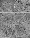
Transmission electron micrographs of the merogonic and early sporogonic stages of Nucleospora hippocampi n. sp. in the nuclei of intestinal epithelial cells of the Hippocampus erectus. (A) Early uninucleate meront stage within the nucleoplasm of the host nucleus. Host nuclear envelope (NE), host mitochondria (M), the monokaryon (n), and plasma membrane (pm) of the meront. (B) The uninucleate meront undergoing karyokinesis. Long microtubules (MT) radiate from two spindle plaques (black arrows). Inset showing the details of the microtubules and spindle plaque. Insert scale bar = 0.1 μm. (C) The binucleate meront contains two nuclei (n) separated by claviform cytoplasm (asterisk). (D) The trinucleate meront with three nuclei and claviform cytoplasm (asterisk). (E) The electron-dense discs (EDD) appear in the cytoplasm of late trinucleate meront (or early sporogonial plasmodium). (F) An early sporogonial plasmodium with increasing EDD around three nuclei. The spindle plaque (black arrow) forming on one nucleus indicates nucleus division again.
Figure 5
Figure 5
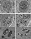
Transmission electron micrographs of the sporogonic stages of Nucleospora hippocampi n. sp. in nuclei of intestinal epithelial cells of Hippocampus erectus. (A) The sporogonial plasmodium contains numerous electron-dense discs (EDD) and four nuclei (n) distributed close to the inner edge of the cytomembrane. (B) Multinucleate sporogonial plasmodium contains four sets of the primordial polar tube (white arrows). EDD is associated with primordial polar tube formation. (C) Late-stage sporogonial plasmodium with increased precursors of the polar tube (white arrows) and reduced EDD. The EDD transform into rounded electron-dense bodies (EDB), then the EDB coalesce into primordial turns of the polar tube (black arrow). (D) The sporoblasts (five are visible) develop in direct contact with the host nucleoplasm. The sporoblasts showing parasite nucleus (n), coiled polar tube with 6–7 turns (PF), and thickened plasmalemma (arrowhead). Insert showing the magnification of anchoring disk (AD). Insert scale bar = 0.1 μm. (E) Late immature spore with preformed winding spore wall (white arrowhead), posterior vacuole (PV), and polar tube (PF). (F) Mature spore with smooth spore wall (white arrowhead) and thickening electron lucent endospore (EN).
Figure 6
Figure 6
Transmission electron micrographs showing the ultrastructure of mature spores of Nucleospora hippocampi n. sp. in the Hippocampus erectus. (A–C) Three spores showing the typical microsporidian structures and organelles. The spore wall (W), anchoring disk (AD), polaroplast (PP), posterior vacuole (PV), polar filament (PF), and the single nucleus (n) are indicated. (D) Ultrastructural detail of spore wall showing the exospore (black arrow), the endospore (asterisk), and the plasmalemma (arrowhead). Notice the electron-dense materials deposited on the outer surface of the exospore (white arrow). (E) Detail of terminal anchoring disk and associated polar tube (PF). (F) High magnification of a transverse section of a spore showing the lamellar region of the polaroplast (PP). (G) The single nucleus (n) is situated beside the posterior vacuole (PV). (H) Detail of the isofilar polar filament (PF).
Figure 7
Figure 7
The Maximum likelihood and Bayesian analyses based on SSU rDNA sequences showing the relationship of Nucleospora hippocampi n. sp. and other selected microsporidian species. Parahepatospora carcini (KX757849), Microsporidium sp. MIC2 (FJ794867), and Microsporidium sp. DP-1-19 (AF394528) are used as the out-group. Values at nodes represent Maximum-likelihood bootstrap support percentages (BP)/Bayesian posterior probabilities (PP). The scale bar represents the estimated number of substitutions per nucleotide site.
Figure 8
Figure 8
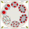
Diagrammatic representation of the proposed intranuclear life cycle of Nucleospora hippocampi n. sp. (1) Uninucleate meront free in the host nucleus. (2) Binucleate meront formed by nucleus mitosis without cytokinesis. (3) The nuclear divides again to produce a tetra-nucleate meront with numerous claviform cytoplasm. (4) The proliferative sporogonial plasmodium shows electron-dense disks and eight units of polar tube precursors around the inner edge of its plasmalemma. (5) The syncytial sporogonial plasmodium produces eight uninucleate sporoblasts by rosette-like budding. The invagination of plasmalemma segregates each nucleus with polar tube precursors into each sporoblast. (6) The individual sporoblasts develop into immature spores. (7) Eight mature spores are formed in direct contact with the host nucleoplasm.

Similar articles

Cited by

References

    1. Balbiani E. G. (1882). Sur les Microsporidies ou Psorospermies des Articules. Compt. Rend. Acad. Sci. 95, 1168–1171.
    1. Bass D., Czech L., Williams B. A. P., Berney C., Dunthorn M., Mahé F., et al. . (2018). Clarifying the Relationships Between Microsporidia and Cryptomycota. J. Eukaryot. Microbiol. 65 (6), 773–782. doi: 10.1111/jeu.12519 - DOI - PMC - PubMed
    1. Blasiola J. G. (1979). Glugea Heraldi N. Sp. (Microsporida, Glugeidae) From the Seahorse Hippocampus Erectus Perry. J. Fish Dis. 2 (6), 493–500. doi: 10.1111/j.1365-2761.1979.tb00410.x - DOI
    1. Cali A., Owen R. L. (1990). Intracellular Development of Enterocytozoon, a Unique Microsporidian Found in the Intestine of AIDS Patients. J. Protozool. 37 (2), 145–155 doi: 10.1111/j.1550-7408.1990.tb05885.x - DOI - PubMed
    1. Cavalier-Smith T. (1987). The Origin of Eukaryote and Archaebacterial Cells. Ann. N. Y. Acad. Sci. 503 (1), 17–54. doi: 10.1111/j.1749-6632.1987.tb40596.x - DOI - PubMed

Publication types

LinkOut - more resources