Review
. 2022 May 18;3(2):e144.
doi: 10.1002/mco2.144. eCollection 2022 Jun.

Epithelial-mesenchymal transition: The history, regulatory mechanism, and cancer therapeutic opportunities

Affiliations
Review

Epithelial-mesenchymal transition: The history, regulatory mechanism, and cancer therapeutic opportunities

Zhao Huang et al. MedComm (2020). .

Abstract

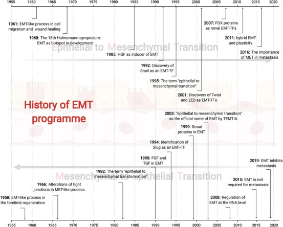
Epithelial-mesenchymal transition (EMT) is a program wherein epithelial cells lose their junctions and polarity while acquiring mesenchymal properties and invasive ability. Originally defined as an embryogenesis event, EMT has been recognized as a crucial process in tumor progression. During EMT, cell-cell junctions and cell-matrix attachments are disrupted, and the cytoskeleton is remodeled to enhance mobility of cells. This transition of phenotype is largely driven by a group of key transcription factors, typically Snail, Twist, and ZEB, through epigenetic repression of epithelial markers, transcriptional activation of matrix metalloproteinases, and reorganization of cytoskeleton. Mechanistically, EMT is orchestrated by multiple pathways, especially those involved in embryogenesis such as TGFβ, Wnt, Hedgehog, and Hippo, suggesting EMT as an intrinsic link between embryonic development and cancer progression. In addition, redox signaling has also emerged as critical EMT modulator. EMT confers cancer cells with increased metastatic potential and drug resistant capacity, which accounts for tumor recurrence in most clinic cases. Thus, targeting EMT can be a therapeutic option providing a chance of cure for cancer patients. Here, we introduce a brief history of EMT and summarize recent advances in understanding EMT mechanisms, as well as highlighting the therapeutic opportunities by targeting EMT in cancer treatment.

Keywords: EMT; cancer progression; embryogenesis; redox signaling; targeted therapy.


Conflict of interest statement

Canhua Huang is an editorial board member of MedComm. Author Canhua Huang was not involved in the journal's review of, or decisions related to, this manuscript. The other authors have no conflicts of interest to declare.

Figures

FIGURE 1
FIGURE 1
A brief history of EMT. The EMT phenotype was firstly discovered by Elizabeth D Hay, who is the pioneer for this field. From 1950s to1980s, EMT was largely investigated in the context of developmental biology with few mechanistic studies. During 1980s to 2010, the connection between EMT and cancer metastasis was intensively documented, and mechanistic studies revealed the central roles of EMT‐TFs in EMT program. From 2010s to now, new concepts of EMT is increasingly developed, which include novel EMT regulators, the hybrid EMT state, multiple cancer hallmarks induced by EMT, the tumor‐suppressive effects of EMT, and EMT‐based cancer therapeutic strategies
FIGURE 2
FIGURE 2
An overview of cellular phenotype changes during EMT. EMT is a highly reversible process with epithelial, hybrid, and mesenchymal states. In epithelial state, cells are hold together to preserve epithelial integrity via several junction structures, namely adherens junctions, tight junctions, desmosomes, and gap junctions, which is composed of several epithelial proteins including E‐cadherin, claudins, occludins, connexins, and many others. Disruption of these junctions leads to the entry of cancer cells into hybrid state and following mesenchymal state, in which cells express mesenchymal marks such as N‐cadherin, Vimentin, and MMPs. In addition, cells reorganize their cytoskeleton networks to support the formation of pseudopodia thereby facilitating metastasis
FIGURE 3
FIGURE 3
EMT‐TFs are key drivers of EMT program. The EMT program is controlled by several EMT‐TFs, including Snail, Twist, and ZEB. These EMT‐TFs are regulated at transcriptional and posttranslational levels, such as protein phosphorylation, ubiquitination, and RNA m6A modification. The m6A modification on EMT‐TF mRNAs can be recognized by different m6A readers, which facilitate the translation or promote RNA decay. The protein phosphorylation is coordinated by kinases and phosphatases, whereas the ubiquitination can be balanced via E3 ligases and deubiquitinases. When translocated into nucleus, EMT‐TFs bind with different epigenetic modifiers such as EZH2, HDAC1/2, BMI1 to form transcriptional complexes, thereby regulating EMT program
FIGURE 4
FIGURE 4
The crosstalk between EMT and TGFβ signaling. Activated TGFβ signaling leads to the nuclear translocation of Smad2/3/4 complex, which directly recognizes the promoters of EMT‐TFs to initiate their transcription. In turn, EMT‐TFs can bind with Smad proteins to form EMT‐promoting Smad complex (EPSC), thus regulating the transcription of epithelial and mesenchymal marks. Besides, TGFβ signaling also regulate the expression of EMT‐TFs through noncoding RNAs
FIGURE 5
FIGURE 5
Regulation of EMT via canonical or noncanonical Wnt pathway. In canonical Wnt pathway, nuclear β‐catenin/LEF/TCF transcriptional complex activates the transcription of EMT‐TFs, thus regulating EMT. In turn, EMT‐TFs affect Wnt signaling to form feedback loops. For example, Snail/β‐catenin complex activates transcription of TCF1, which is the key transcription factor of Wnt signaling. ZEB represses the transcription of miR‐200a, leading to the reactivation of β‐catenin. Twist can transcribe Wnt5a, which activates Wnt receptor Frizzled. In noncanonical Wnt pathway, activation of Frizzled2 leads to the phosphorylation and consequent nuclear translocation of STAT3, which upregulates mesenchymal marks and represses epithelial marks independent of β‐catenin
FIGURE 6
FIGURE 6
Hedgehog signaling regulates EMT and cancer progression. Hedgehog (Hh)‐mediated inhibition of PTCH1 leads to the activation of SMO, which promotes the activation of the transcription factor Gli1/2. The target genes of Gli1/2 include several EMT associated proteins such as Snail and E‐cadherin. Besides, Snail and Gli share the same protein turnover system, namely β‐TrCP‐mediated protein degradation and USP37‐induced protein stabilization. In turn, cellular EMT status affects Hedgehog signaling, as evidenced by loss of E‐cadherin has been shown to activate Gli1/2
FIGURE 7
FIGURE 7
The connection between EMT and Hippo signaling. When Hippo signaling is inactivated, YAP and TEAD form complex in nucleus to direct transcription. EMT‐TFs can bind with YAP/TEAD complex, leading to a functional switch of EMT‐TFs from epigenetic repressors into activators, resulting in the overexpression of YAP target genes to facilitate tumor progression. Besides, Hippo signaling can regulate EMT via the interplay with other developmental pathways, such as TGFβ signaling and Wnt signaling. For instance, YAP can stabilize Smad3, the component of TGFβ signaling, thus promoting TGFβ‐mediated EMT. Besides, YAP/TEAD complex can be associated with β‐catenin to form a novel transcriptional complex, which activates the transcription of EMT‐TFs. Moreover, TEAD4 can transcriptionally activate the mesenchymal protein vimentin independent of YAP
FIGURE 8
FIGURE 8
Regulation of EMT by oxidative stress and redox signaling. Redox signaling modulates EMT program via the regulation of redox sensors, including AP‐1, PHD, IKKγ, FOXO1, and many others. Briefly, these redox sensors can be either activated or inactivated via cysteine oxidation, thereby positively or negatively regulate the transcription of EMT‐TFs. Moreover, FOXO1 mRNA acts as ceRNA which binds with miR‐9, protecting E‐cadherin mRNA from miR‐9‐mediated degradation. This effect inhibits EMT program independent of EMT‐TFs

Similar articles

Cited by

References

    1. Yang J, Antin P, Berx G, et al. Guidelines and definitions for research on epithelial‐mesenchymal transition. Nat Rev Mol Cell Biol. 2020;21(6):341‐352. - PMC - PubMed
    1. Dongre A, Weinberg RA. New insights into the mechanisms of epithelial‐mesenchymal transition and implications for cancer. Nat Rev Mol Cell Biol. 2019;20(2):69‐84. - PubMed
    1. Gonzalez DM, Medici D. Signaling mechanisms of the epithelial‐mesenchymal transition. Sci Signal. 2014;7(344):re8. - PMC - PubMed
    1. Caramel J, Papadogeorgakis E, Hill L, et al. A switch in the expression of embryonic EMT‐inducers drives the development of malignant melanoma. Cancer Cell. 2013;24(4):466‐480. - PubMed
    1. Fazilaty H, Rago L, Kass Youssef K, et al. A gene regulatory network to control EMT programs in development and disease. Nat Commun. 2019;10(1):5115. - PMC - PubMed