. 2022 Feb 18;7(7):e202103903.
doi: 10.1002/slct.202103903. Epub 2022 Feb 17.

Identification of a Potential mRNA-based Vaccine Candidate against the SARS-CoV-2 Spike Glycoprotein: A Reverse Vaccinology Approach

Affiliations

Identification of a Potential mRNA-based Vaccine Candidate against the SARS-CoV-2 Spike Glycoprotein: A Reverse Vaccinology Approach

Olanrewaju Ayodeji Durojaye et al. ChemistrySelect. .

Abstract

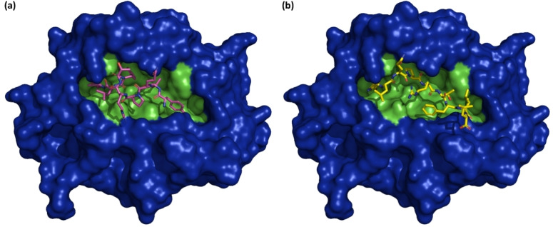
The emergence of the novel coronavirus (SARS-CoV-2) in December 2019 has generated a devastating global consequence which makes the development of a rapidly deployable, effective and safe vaccine candidate an imminent global health priority. The design of most vaccine candidates has been directed at the induction of antibody responses against the trimeric spike glycoprotein of SARS-CoV-2, a class I fusion protein that aids ACE2 (angiotensin-converting enzyme 2) receptor binding. A variety of formulations and vaccinology approaches are being pursued for targeting the spike glycoprotein, including simian and human replication-defective adenoviral vaccines, subunit protein vaccines, nucleic acid vaccines and whole-inactivated SARS-CoV-2. Here, we directed a reverse vaccinology approach towards the design of a nucleic acid (mRNA-based) vaccine candidate. The "YLQPRTFLL" peptide sequence (position 269-277) which was predicted to be a B cell epitope and likewise a strong binder of the HLA*A-0201 was selected for the design of the vaccine candidate, having satisfied series of antigenicity assessments. Through the codon optimization protocol, the nucleotide sequence for the vaccine candidate design was generated and targeted at the human toll-like receptor 7 (TLR7). Bioinformatics analyses showed that the sequence "UACCUGCAGCCGCGUACCUUCCUGCUG" exhibited a strong affinity and likewise was bound to a stable cavity in the TLR7 pocket. This study is therefore expected to contribute to the research efforts directed at securing definitive preventive measures against the SARS-CoV-2 infection.

Keywords: Antigens; Reverse vaccinology; SAR-CoV-2; Spike glycoprotein; Vaccine; Viruses.


Conflict of interest statement

The authors declare no conflict of interest.

Figures

Figure 1
Figure 1
ProPred‐I output for promiscuous MHC Class‐I binding Peptides. The server represents predicted binders using color variations. The predicted binders are represented in blue color while the red‐colored peptides represent the first position of each binder, for easy identification of overlapping peptides.
Figure 2
Figure 2
(a) BepiPred‐2.0 B cell epitope prediction for the SARS‐CoV‐2 spike glycoprotein. The “Epitopes” row indicates the position of residues with scores above the set epitope threshold. “Predictions” illustrate the predictions of BepiPred‐2.0 with the amino acid residues of the protein displayed in orange gradient. “Structural” illustrate the probability gradient of the alpha helix (H) (which is not covered in the figure), beta sheet (E) with blue gradient, and coils (C), with orange gradient. The exposed (E) and buried (B) regions of the sequence are illustrated by the “Surface” column which was obtained using the NetsurfP default threshold. The orange gradient in this column illustrates the predicted relative surface accessibility. (b) An illustration of the SARS‐CoV‐2 surface representation (PDB 6VXX), with the predicted antigenic region highlighted in red color.
Figure 3
Figure 3
BcePred graphical output for the SARS‐CoV‐2 spike glycprotein B cell epitope prediction. The displayed graph covers the amino acid residues between 241 to 300 of the viral sequence.
Figure 4
Figure 4
TMHMM graphical display of the SARS‐CoV‐2 spike glycoprotein transmembrane propensity prediction. The vertical red lines of the plot illustrate the predicted transmembrane helical segments of the protein. The blue line depicts the probability of a segment of the protein sequence to be intracellular while the outer membrane segment is denoted in pink line.
Figure 5
Figure 5
(a) TOPCONS predicted transmembrane topology with the predicted ΔG values. Transmembrane helices as denoted in the keys shown in the figure, are the grey and white rectangles. (b) Consensus prediction of the TOPCONS, detecting short sequence regions of the protein as predicted signal peptides (vertical black rectangle) and transmembrane helices (vertical white rectangle) respectively.
Figure 6
Figure 6
Predicted N‐glycosylation sites in the SARS‐CoV‐2 spike glycoprotein.
Figure 7
Figure 7
Stick representation of the top five generated models from the CABS‐flex simulation protocol.
Figure 8
Figure 8
Binding poses for the peptide model with the strongest binding energy (model 2) in the HLA*A‐0201 binding pocket as obtained from (a) the ClusPro model (purple sticks) and (b) the HPEPDOCK peptide binding model (yellow sticks).
Figure 9
Figure 9
Interaction analysis of HLA*A‐0201‐peptide complex in 3D and 2D representations. (a) The HPEPDOCK‐derived docked HLA*A‐0201‐peptide complex used as the starting complex for the molecular dynamics simulation. (b) A representative snapshot of the simulated HLA*A‐0201‐peptide complex.
Figure 10
Figure 10
Molecular dynamics simulation and trajectory analysis of the free HLA*A*0102 and HLA*A*0102‐peptide complex. Plots of the (a) Root‐mean‐square deviation, (b) Root‐mean‐square fluctuation, (c) Radius of gyration, (d) Solvent‐accessible surface area, (e) Intramolecular hydrogen bonding, and (f) principal component analyses.
Figure 11
Figure 11
Amino acids colored based on the change in vibrational entropy upon mutation. Blue color signifies structural rigidification while red signifies flexibility gain. The output (blue colored) is an indication of an increase in stability upon the N501Y mutation on the SARS‐CoV‐2 spike glycoprotein.
Figure 12
Figure 12
Interatomic interaction prediction upon the N501Y mutation. Residues of the wild‐type (a) and mutant (b) are colored in light‐green and are also presented along with the surrounding residues as sticks. The surrounding residues are involved in other types of interaction. The bonds are colored according to interaction types. The red, yellow and pink represent hydrogen bonds, ionic interactions and carbonyl contacts respectively.
Figure 13
Figure 13
Visual analysis of deformation energies and atomic fluctuation. The deformation magnitude is illustrated by thin to thick colored tubes. Blue white and red represent the low, moderate and high magnitudes respectively (a1 and b1). The fluctuation magnitude is also illustrated by thin to thick colored tubes, where the blue white and red represent the low, moderate and high magnitudes respectively (a2 and b2).
Figure 14
Figure 14
Secondary and tertiary structure depiction of the top five SimRNA simulation output. For each cluster, the tertiary structures are displayed in their 3D format above the corresponding secondary structures.
Figure 15
Figure 15
Cartoon representation of the binding pose of “Cluster 1” mRNA vaccine candidate in the human TLR7 binding pocket (a). Surface representation of the human TLR7 is displayed, with meshes in different color gradients denoting the different degrees of stability. Green mesh denotes a metastable pocket while blue and red denote the unstable and stable pockets respectively (b).
Figure 16
Figure 16
3‐D interaction analysis of the Cluster 1 mRNA with the TLR7 pocket residues.

Similar articles

Cited by

References

    1. Drosten C., Günther S., Preiser W., Van Der Werf S., Brodt H.-R., Becker S., Rabenau H., Panning M., Kolesnikova L., Fouchier R. A., N. Engl. J. Med. 2003, 348 (20), 1967. - PubMed
    1. Zaki A. M., Van Boheemen S., Bestebroer T. M., Osterhaus A. D., Fouchier R. A., N. Engl. J. Med. 2012, 367 (19), 1814. - PubMed
    1. Huang C., Wang Y., Li X., Ren L., Zhao J., Hu Y., Zhang L., Fan G., Xu J., Gu X., The Lancet 2020, 395 (10223), 497. - PMC - PubMed
    1. Zhou P., Yang X.-L., Wang X.-G., Hu B., Zhang L., Zhang W., Si H.-R., Zhu Y., Li B., Huang C.-L., Nature 2020, 579 (7798), 270. - PMC - PubMed
    1. N. Zhu, D. Zhang, W. Wang, X. Li, B. Yang, J. Song, X. Zhao, B. Huang, W. Shi, R. Lu, A novel coronavirus from patients with pneumonia in China, 2019. N. Engl. J. Med. 2020. - PMC - PubMed

LinkOut - more resources