. 2022 May 7:15:100280.
doi: 10.1016/j.mtbio.2022.100280. eCollection 2022 Jun.

Fusing spheroids to aligned μ-tissues in a heart-on-chip featuring oxygen sensing and electrical pacing capabilities

Affiliations

Fusing spheroids to aligned μ-tissues in a heart-on-chip featuring oxygen sensing and electrical pacing capabilities

Oliver Schneider et al. Mater Today Bio. .

Abstract

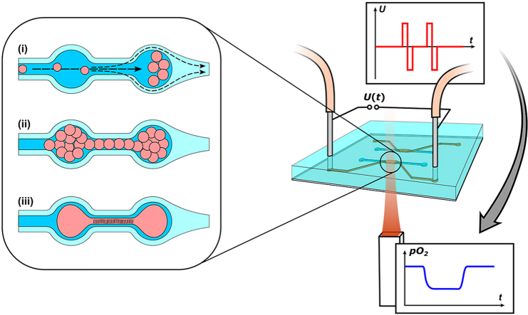
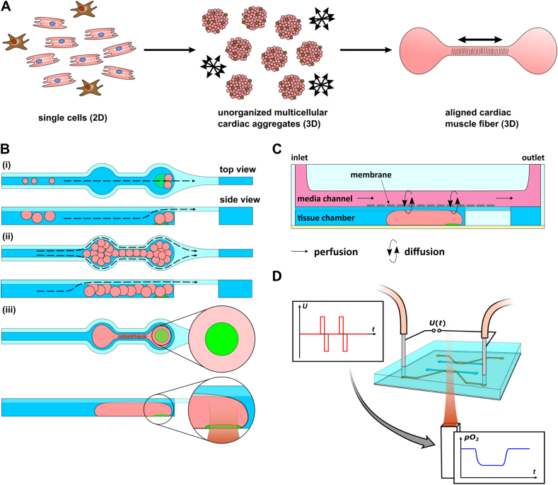
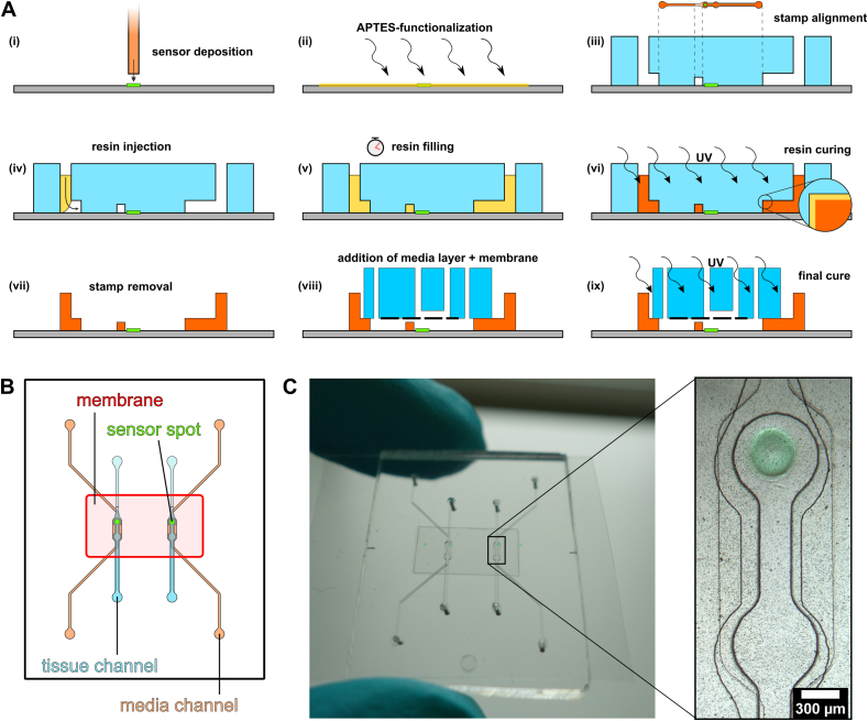
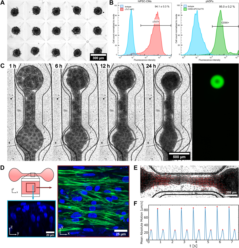
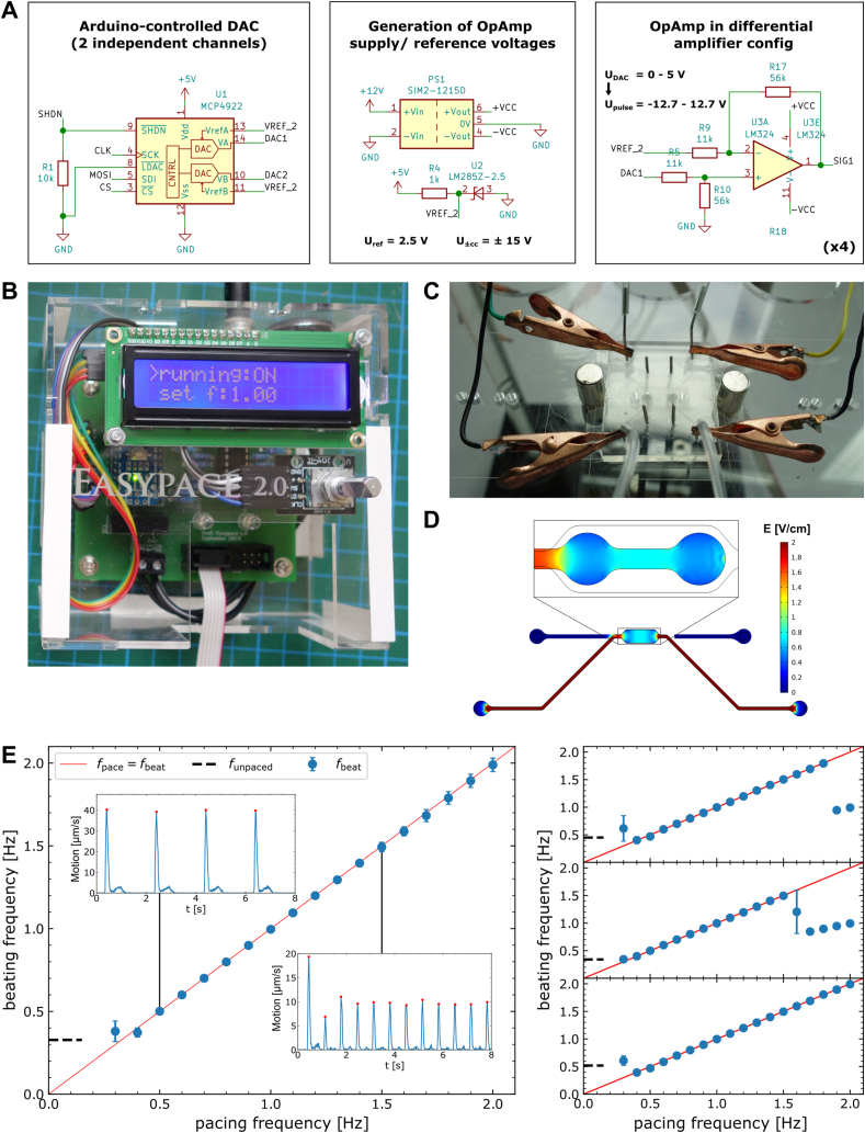
Over the last decade, Organ-on-Chip (OoC) emerged as a promising technology for advanced in vitro models, recapitulating key physiological cues. OoC approaches tailored for cardiac tissue engineering resulted in a variety of platforms, some of which integrate stimulation or probing capabilities. Due to manual handling processes, however, a large-scale standardized and robust tissue generation, applicable in an industrial setting, is still out of reach. Here, we present a novel cell injection and tissue generation concept relying on spheroids, which can be produced in large quantities and uniform size from induced pluripotent stem cell-derived human cardiomyocytes. Hydrostatic flow transports and accumulates spheroids in dogbone-shaped tissue chambers, which subsequently fuse and form aligned, contracting cardiac muscle fibers. Furthermore, we demonstrate electrical stimulation capabilities by utilizing fluidic media connectors as electrodes and provide the blueprint of a low-cost, open-source, scriptable pulse generator. We report on a novel integration strategy of optical O2 sensor spots into resin-based microfluidic systems, enabling in situ determination of O2 partial pressures. Finally, a proof-of-concept demonstrating electrical stimulation combined with in situ monitoring of metabolic activity in cardiac tissues is provided. The developed system thus opens the door for advanced OoCs integrating biophysical stimulation as well as probing capabilities and serves as a blueprint for the facile and robust generation of high density microtissues in microfluidic modules amenable to scaling-up and automation.

Keywords: CMs, Cardiomyocytes; Electrical stimulation; HoC, Heart-on-Chip; Metabolism; Microphysiological systems; Noninvasive readouts; OoC, Organ-on-Chip; Optical sensors; Organ-on-Chip; hiPSCs, human induced pluripotent stem cells.


Conflict of interest statement

The authors declare the following competing interest(s): T.M. is a founder, holds equity in PyroScience GmbH in Germany, and is the CEO of the Austrian branch, PyroScience AT GmbH. PyroScience is a developer, producer, and vendor of sensor technology.

Figures

Image 1
Graphical abstract
Fig. 1
Fig. 1
Concept of the Spheroflow HoC: A) Process for generating aligned cardiac fibers: Initial compaction of single cells of precisely defined mixture to 3D spheroids. Spheroids further merge to an aligned tissue fiber, guided by the chamber geometry. Double headed arrows indicate alignment direction. B) Chip loading mechanism: (i) Spheroids are introduced by hydrostatically driven flow into the dogbone-shaped tissue compartment and confined by the channel constriction. (ii) With gradual filling, the loading flow is maintained by the flanking constriction channel. (iii) Individual spheroids merge to a single aligned cardiac tissue. Embedded O2 sensor in the knob region enables in situ readout of O2 levels. C) Side view of the chip depicting the nutrient supply: A syringe pump generates a defined media flow in the media channels. The porous membrane protects the cultured tissue from excessive shear forces while allowing diffusive transport of nutrients and waste products. D) Investigation of metabolic activity in perfused tissues. Presented platform enables electrical tissue stimulation coupled to optical readout of O2 levels.
Fig. 2
Fig. 2
Microfluidic chip fabrication: A) Microfabrication steps (i-ix): Deposition of O2 sensor onto bottom layer (i) and subsequent surface functionalization (ii). Placement of PDMS stamp on bottom layer precisely aligning the dogbone-knob onto the sensor spot (iii). Injection (iv), capillary filling (v) and curing (vi) of resin. Resin in contact with PDMS does not solidify due to locally available O2 and remains sticky (inset). Removal of PDMS stamp exposes open tissue channels with an uncured resin layer (vii). Addition of APTES-treated media layer with bonded membrane (viii) and another UV curing (ix) finalizes the chip. B) Design of Spheroflow HoC: Each chip comprises two individual systems consisting of a loading and media channel, separated by a porous membrane. O2 sensor spots are integrated into the knob region of the tissue chamber. C) Assembled chip with integrated sensors. Magnified view depicts a micrograph of a dogbone-shaped tissue chamber with integrated O2 sensor spot.
Fig. 3
Fig. 3
Formation of cardiac μ-tissues: A) Generation of uniformly sized spheroids in μ-Wells. B) Flow cytometry analysis of CM and FB cell populations prior to spheroid formation. C) Fusing of individual cardiac spheroids to an aligned tissue over the timespan of 24 ​h after loading. The underlying integrated O2 sensor spot is visualized via widefield fluorescence microscopy (green, Cy5 filterset). D) Characterization of the structure of the generated cardiac muscle fiber in fibronectin-coated HoC by immunostaining of cTnT (green) and DAPI (blue). The shaft region is characterized by aligned fibers (xy slice). The perpendicular z-stack view reveals stacked nuclei, indicating the generation of a multilayered 3D tissue (yz slice). E) Characterization of beating motion of a fiber. Motion vectors at tissue contraction reveal a collective displacement. F) Optically determined beating kinetics display pronounced peaks of contraction (red circles) and relaxation (red triangles), amenable for beating frequency extraction.
Fig. 4
Fig. 4
Electrical stimulation of cardiac tissues: A) Electrical schematic of key components of the pulse generator ‘Easypace’. An Arduino-controlled DAC creates two independent output signals that are subsequently converted to biphasic pulses via an Operational Amplifier. B) Picture of the pulse generator ‘Easypace’ that can be controlled either manually via the rotary encoder or remotely via Serial commands and provides feedback by the integrated LCD display. C) Image of pacing setup. The pulse generator is connected via alligator clips to stainless steel fluidic connectors in the media layer, thus acting as electrodes. D) Simulated electrical field distribution inside media- & tissue-compartments upon application of 10 ​V between media in- & outlet. E) Beating rate analysis of tissues stimulated electrically at increasing frequencies. Beating frequency extracted from beating kinetics (insets) shown as a function of ramped up pacing frequency for four tissues. Ideal matching of beating and pacing frequency indicated by linear relation (red line).
Fig. 5
Fig. 5
In situ measurements of O2 levels: A) Recording of O2 partial pressures of two tissues alternatingly paced (dashed lines) at increasing frequencies of (0.7, 1.0, 1.2, 1.5) Hz. B) Evaluation of frequency dependence of relative jump heights in O2 consumption for tissues repeatedly probed following timings in (A). C) Stability analysis of O2 levels in empty systems perfused with media over 12 ​h. D) Comparison of O2 partial pressures between paced and unpaced chambers, measured in empty, media-perfused systems. No difference between both conditions is observed, excluding O2 changes induced by electrolysis.

Similar articles

Cited by

References

    1. Onakpoya I.J., Heneghan C.J., Aronson J.K. Post-marketing withdrawal of 462 medicinal products because of adverse drug reactions: a systematic review of the world literature. BMC Med. 2016;14:10. doi: 10.1186/s12916-016-0553-2. - DOI - PMC - PubMed
    1. Marsano A., Conficconi C., Lemme M., Occhetta P., Gaudiello E., Votta E., Cerino G., Redaelli A., Rasponi M. Beating heart on a chip: a novel microfluidic platform to generate functional 3D cardiac microtissues. Lab Chip. 2016;16:599–610. doi: 10.1039/C5LC01356A. - DOI - PubMed
    1. Nunes S.S., Miklas J.W., Liu J., Aschar-Sobbi R., Xiao Y., Zhang B., Jiang J., Massé S., Gagliardi M., Hsieh A., Thavandiran N., Laflamme M.A., Nanthakumar K., Gross G.J., Backx P.H., Keller G., Radisic M. Biowire: a platform for maturation of human pluripotent stem cell–derived cardiomyocytes. Nat. Methods. 2013;10:781–787. doi: 10.1038/nmeth.2524. - DOI - PMC - PubMed
    1. Mathur A., Loskill P., Shao K., Huebsch N., Hong S., Marcus S.G., Marks N., Mandegar M., Conklin B.R., Lee L.P., Healy K.E. Human iPSC-based cardiac microphysiological system for drug screening applications. Sci. Rep. 2015;5:8883. doi: 10.1038/srep08883. - DOI - PMC - PubMed
    1. Zhang Y.S., Aleman J., Arneri A., Bersini S., Piraino F., Shin S.R., Dokmeci M.R., Khademhosseini A. From cardiac tissue engineering to heart-on-a-chip: beating challenges, Biomed. Mater. 2015;10 doi: 10.1088/1748-6041/10/3/034006. - DOI - PMC - PubMed