. 2022 May 5;25(6):104355.
doi: 10.1016/j.isci.2022.104355. eCollection 2022 Jun 17.

The Taspase1/Myosin1f-axis regulates filopodia dynamics

Affiliations

The Taspase1/Myosin1f-axis regulates filopodia dynamics

Astrid Hensel et al. iScience. .

Abstract

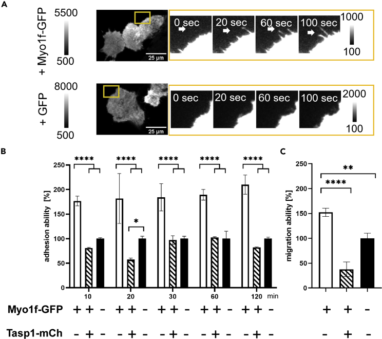
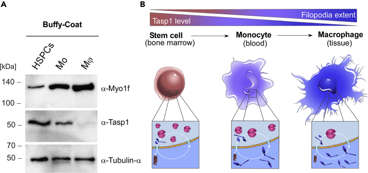
The unique threonine protease Tasp1 impacts not only ordered development and cell proliferation but also pathologies. However, its substrates and the underlying molecular mechanisms remain poorly understood. We demonstrate that the unconventional Myo1f is a Tasp1 substrate and unravel the physiological relevance of this proteolysis. We classify Myo1f as a nucleo-cytoplasmic shuttle protein, allowing its unhindered processing by nuclear Tasp1 and an association with chromatin. Moreover, we show that Myo1f induces filopodia resulting in increased cellular adhesion and migration. Importantly, filopodia formation was antagonized by Tasp1-mediated proteolysis, supported by an inverse correlation between Myo1f concentration and Tasp1 expression level. The Tasp1/Myo1f-axis might be relevant in human hematopoiesis as reduced Tasp1 expression coincided with increased Myo1f concentrations and filopodia in macrophages compared to monocytes and vice versa. In sum, we discovered Tasp1-mediated proteolysis of Myo1f as a mechanism to fine-tune filopodia formation, inter alia relevant for cells of the immune system.

Keywords: Biological sciences; Cell biology; Immunology.


Conflict of interest statement

The authors declare no competing interests.

Figures

None
Graphical abstract
Figure 1
Figure 1
Myo1f is a bona fide substrate of Tasp1 (A/B) A semi-in vitro Tasp1 substrate cleavage assay was employed to verify the predicted Tasp1 cleavage site of Myo1f. 293T cells were transfected with plasmids encoding Myo1f-GFP or the cleavage site-deficient mutant Myo1f-245AA246-GFP. 24 h after transfection, cell lysates were incubated with or without 10 μM recombinant Tasp1-His at 37°C for the indicated time periods. The assay was performed without (A) or with (B) a two-fold concentrated protease inhibitor mix. Immunoblot analysis revealed the appearance of the C-terminal cleavage product (calculated molecular weight: 124 kDa) only in case of wild-type Myo1f and Tasp1-His presence confirming a Tasp1-His mediated cleavage of full-length (FL) Myo1f-GFP. (C) Schematic tripartite structure of Myo1f comprising a head (blue oval), a neck (blue sphere) and a tail (gray) domain. Indicated are the proposed nuclear localization signal (NLS) and the Tasp1 cleavage site (CS), the ATP and actin binding domains located within the head domain, and the nuclear export signal (NES)-bearing region (red) arranged between the IQ calmodulin-binding motif, the motor and tail homology 1 and 2 (TH1/TH2) and src homology 3 domains as part of the tail. (D) 293T cells co-expressing the indicated Myo1f variants and Tasp1-HA were treated with 50 nM bortezomib. Immunoblotting of whole cell extracts revealed a lower migrating band only for WT Myo1f-GFP after proteasome inhibition.
Figure 2
Figure 2
Myo1f is a nucleo-cytoplasmic shuttle protein (A) Schematic presentation of full-length Myo1f-GFP (left) and myc-/GFP-labeled Myo1f-truncations representing the N- (middle) and the C-terminal Tasp1 cleavage product (right). (B) Confocal fluorescence microscopy images showing the subcellular localization of Myo1f and truncated variants in HeLa cells. Full length Myo1f-GFP exhibited a predominantly cytosolic localization, but exposure to Leptomycin B (LMB) results in a shift to an increased nuclear localization (left panel). In contrast, the N-terminal fragment clearly localized to the nucleus, whereas the C-terminal fragment was exclusively cytoplasmic (right panel). Scale bars 25 μm. (C) Quantification of the intracellular localization of full-length Myo1f-GFP using the software “CellProfiler3” (Schindelin et al., 2012). Mean nuclear fluorescence intensity between 500 nm and 550 nm was measured in ROIs defined by Hoechst staining. Error bars represent the CI 95%, asterisks indicate statistical significance of mean difference assessed by t-test (∗: p ≤ 0.05; ∗∗: p ≤ 0.01; ∗∗∗: p ≤ 0.001). (D) Gaussian fit of the frequency distributions of measured mean nuclear fluorescence intensities further substantiated the increased nuclear localization upon LMB treatment, as two distinct populations become apparent. n = 2,572 cells from 4 independent experiments. (E) Myo1f-GFP and Myo1f-245AA246-GFP interact with RNA polymerase II and Histone H3. Co-immunoprecipitation of GFP-tagged Myo1f-variants from 293T chromatin fractions as detected by immunoblot analysis. Input = chromatin fraction; eluate, 40× concentrated. See also Figures S1 and S2.
Figure 3
Figure 3
Myo1f-GFP induces filopodia-like membrane protrusions (A) Myo1f-GFP expressing HeLa cells are characterized by increased filopodia formation as detected by spinning disc confocal microscopy. Scale bars 25 μm. (B) FiloQuant analysis of relative filopodia density (filopodia number per μm cell surface) in HeLa cells expressing Myo1f-GFP compared to GFP expressing control cells. Statistical significance was assessed by t-test with a total of n = 100 cells/group in 3 independent experiments (∗: p < 0,05). Error bars represent the standard deviation. (C) FiloQuant measurement of filopodia longer than 1 μm in HeLa cells expressing Myo1f-GFP compared to GFP expressing control cells. n = 100 cells/group from 3 independent experiments. (D) Formation of Myo1f-GFP-induced cell protrusions containing filamentous F-actin in different adherent cell lines. 293T-, HeLa- and SW480 cells were transfected with either Myo1f-GFP (left panel) or GFP (right panel) and fixed 24 h later. F-actin was stained with rhodamine-conjugated phalloidin (red) and cells were analyzed by laser scanning confocal microscopy. Scale bars 25 μm. (E) THP-1 monocytes (Mo) were differentiated into macrophage-like cells (THP-1 macrophages, Mφ) by PMA treatment and analyzed by immunostaining. Endogenous Myo1f of THP-1 macrophages localizes to cell protrusions together with filamentous actin whereas THP-1 monocytes show comparatively less endogenous Myo1f and no filopodia. Scale bars 25 µm. (F) Immunoblot showing varying Tasp1 expression in different cell lines. (G) Decreased Tasp1 expression coincides with increased full-length Myo1f protein levels during differentiation from monocytes (Mo) into macrophages (Mφ) as demonstrated by immunoblot analysis. See also Figures S3 and S4.
Figure 4
Figure 4
Tasp1 co-expression reduces filopodia in cells expressing Myo1f but not in cells expressing a non-cleavable Myo1f mutant (A/B) Counting of filopodia longer than 5 μm in HeLa (A) and 293T (B) cells expressing Myo1f- or Myo1f-245AA246-GFP together with either Tasp1-mCherrry (n = 83/n = 87) or a mock plasmid (n = 114/n = 94); filopodia counts per cell were normalized to the respective control cells without Tasp1 co-expression. Significance was analyzed by an unpaired t-test (∗∗: p ≤ 0.01; ns: not significant). Error bars indicate the standard deviation. (C) Representative confocal microscopy images of cells expressing the indicated constructs. Scale bars 25 μm.
Figure 5
Figure 5
Myo1f facilitates cellular adhesion and migration (A) Myo1f-induced filopodia actively sense their environment. Representative TIRF images of cells overexpressing Myo1f-GFP or GFP control. Yellow box indicates zoomed time-lapse sequential TIRF images which show a dynamic alternation of filopodia growth and retraction as indicated by the white arrows. Scale bars 25 μm. (B) Overexpression of Myo1f-GFP enhances the adhesion of 293T cells on fibronectin-coated plates as assessed by a MTS assay at the indicated time points. This effect was significantly counteracted by co-expression of Tasp1-mCherry. Error bars show the SD (n = 3), asterisks indicate significant differences in mean comparison by two-Way ANOVA(∗: p ≤ 0.05; ∗∗∗∗: p ≤ 0.0001). (C) Overexpression of Myo1f-GFP significantly increases the migration capacity of 293T cells as assessed by a Boyden chamber assay. Error bars show the SD (n = 3), asterisks indicate significant differences evaluated by two-Way ANOVA (∗∗: p ≤ 0.01, ∗∗∗∗: p ≤ 0.001).
Figure 6
Figure 6
The Myo1f/Tasp1-axis enables regulation of filopodia formation during immune cell differentiation (A) Reduced Tasp1 expression coincides with increased full-length Myo1f concentration during the differentiation of hematopoietic stem and myeloid progenitor cells to monocytes and finally into macrophages. Hematopoietic stem and progenitor cells (HSPCs) and monocytes (Mo) were isolated from human buffy coats. Moreover, isolated monocytes were differentiated into macrophages (Mφ) and the different immune cell fractions were analyzed by immunoblot, α-Tubulin served as a loading control. (B) Model of Myo1f/Tasp1-regulated filopodia formation during macrophage development: filopodia formation increases during hematopoiesis, while Tasp1 levels decline with a higher state of cellular differentiation. Hematopoietic stem and progenitor cells (HSPCs) in the bone marrow contain high Tasp1 levels, resulting in Myo1f-cleavage and degradation. This in turn might suppress Myo1f-induced filopodia formation and associated migration properties (left). Monocytes have emigrated from the bone marrow into the peripheral blood upon appropriate stimuli. A reduction in Tasp1 concentration allows the increase of Myo1f-provoked filopodia (middle). Transendothelial migration from the circulation into tissues promotes monocyte differentiation into macrophages. The characteristic filopodia phenotype of activated macrophages might be facilitated by uncleaved Myo1f (right). See also Figure S5.

References

    1. Adams J., Palombella V.J., Sausville E.A., Johnson J., Destree A., Lazarus D.D., Maas J., Pien C.S., Prakash S., Elliott P.J. Proteasome inhibitors: a novel class of potent and effective antitumor agents. Cancer Res. 1999;59:2615–2622. - PubMed
    1. Balkin D.M., Poranki M., Forester C.M., Dorsey M.J., Slavotinek A., Pomerantz J.H. TASP1 mutation in a female with craniofacial anomalies, anterior segment dysgenesis, congenital immunodeficiency and macrocytic anemia. Mol. Genet. Genomic Med. 2019;7:e818. doi: 10.1002/mgg3.818. - DOI - PMC - PubMed
    1. Berg J.S., Cheney R.E. Myosin-X is an unconventional myosin that undergoes intrafilopodial motility. Nat. Cell Biol. 2002;4:246–250. doi: 10.1038/ncb762. - DOI - PubMed
    1. Berg J.S., Derfler B.H., Pennisi C.M., Corey D.P., Cheney R.E. Myosin-X, a novel myosin with pleckstrin homology domains, associates with regions of dynamic actin. J. Cell Sci. 2000;113(Pt 19):3439–3451. doi: 10.1242/jcs.113.19.3439. - DOI - PubMed
    1. Bier C., Knauer S.K., Docter D., Schneider G., Krämer O.H., Stauber R.H. The importin-alpha/nucleophosmin switch controls taspase1 protease function. Traffic. 2011;12:703–714. doi: 10.1111/j.1600-0854.2011.01191.x. - DOI - PubMed

LinkOut - more resources