Distinguishing COVID-19 From Influenza Pneumonia in the Early Stage Through CT Imaging and Clinical Features
- PMID: 35602019
- PMCID: PMC9120763
- DOI: 10.3389/fmicb.2022.847836
Distinguishing COVID-19 From Influenza Pneumonia in the Early Stage Through CT Imaging and Clinical Features
Abstract
Background: Both coronavirus disease 2019 (COVID-19) and influenza pneumonia are highly contagious and present with similar symptoms. We aimed to identify differences in CT imaging and clinical features between COVID-19 and influenza pneumonia in the early stage and to identify the most valuable features in the differential diagnosis.
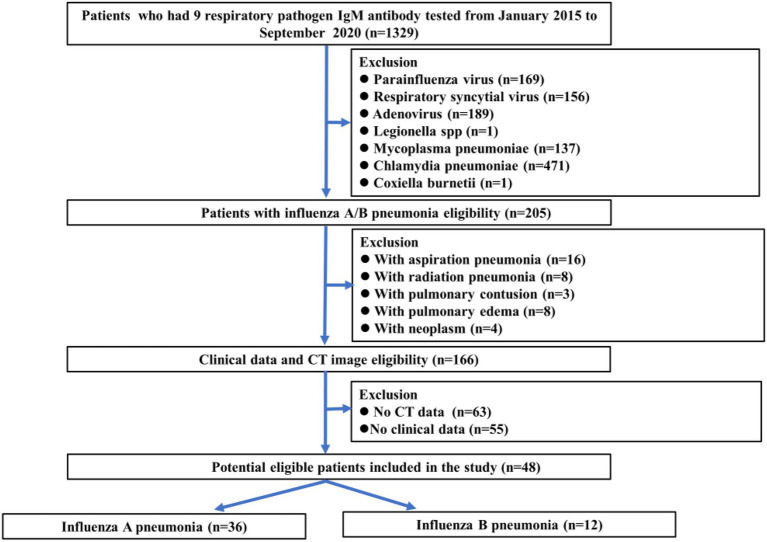
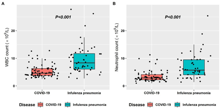
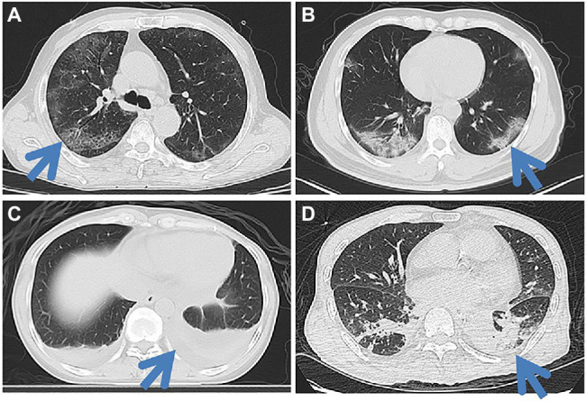
Methods: Seventy-three patients with COVID-19 confirmed by real-time reverse transcription-polymerase chain reaction (RT-PCR) and 48 patients with influenza pneumonia confirmed by direct/indirect immunofluorescence antibody staining or RT-PCR were retrospectively reviewed. Clinical data including course of disease, age, sex, body temperature, clinical symptoms, total white blood cell (WBC) count, lymphocyte count, lymphocyte ratio, neutrophil count, neutrophil ratio, and C-reactive protein, as well as 22 qualitative and 25 numerical imaging features from non-contrast-enhanced chest CT images were obtained and compared between the COVID-19 and influenza pneumonia groups. Correlation tests between feature metrics and diagnosis outcomes were assessed. The diagnostic performance of each feature in differentiating COVID-19 from influenza pneumonia was also evaluated.
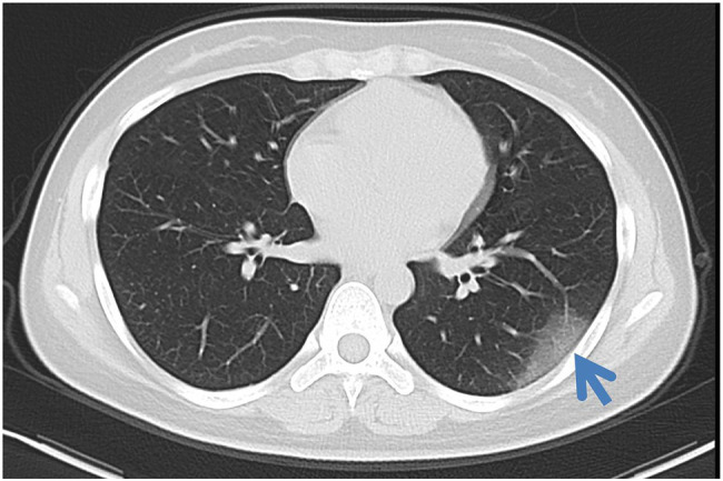
Results: Seventy-three COVID-19 patients including 41 male and 32 female with mean age of 41.9 ± 14.1 and 48 influenza pneumonia patients including 30 male and 18 female with mean age of 40.4 ± 27.3 were reviewed. Temperature, WBC count, crazy paving pattern, pure GGO in peripheral area, pure GGO, lesion sizes (1-3 cm), emphysema, and pleural traction were significantly independent associated with COVID-19. The AUC of clinical-based model on the combination of temperature and WBC count is 0.880 (95% CI: 0.819-0.940). The AUC of radiological-based model on the combination of crazy paving pattern, pure GGO in peripheral area, pure GGO, lesion sizes (1-3 cm), emphysema, and pleural traction is 0.957 (95% CI: 0.924-0.989). The AUC of combined model based on the combination of clinical and radiological is 0.991 (95% CI: 0.980-0.999).
Conclusion: COVID-19 can be distinguished from influenza pneumonia based on CT imaging and clinical features, with the highest AUC of 0.991, of which crazy-paving pattern and WBC count play most important role in the differential diagnosis.
Keywords: COVID-19; CT features; clinical features; differential diagnosis; influenza pneumonia.
Copyright © 2022 Yang, Lin, Chen, Qiu, Li, Huang, Yang, Sun, Liao, Xiao, Tang, Chen, Zhang and Dai.
Conflict of interest statement
The authors declare that the research was conducted in the absence of any commercial or financial relationships that could be construed as a potential conflict of interest.
Figures


Similar articles
-
Temporal relationship between serial RT-PCR results and serial chest CT imaging, and serial CT changes in coronavirus 2019 (COVID-19) pneumonia: a descriptive study of 155 cases in China.Eur Radiol. 2021 Mar;31(3):1175-1184. doi: 10.1007/s00330-020-07268-9. Epub 2020 Sep 15. Eur Radiol. 2021. PMID: 32930834 Free PMC article.
-
Combining initial chest CT with clinical variables in differentiating coronavirus disease 2019 (COVID-19) pneumonia from influenza pneumonia.Sci Rep. 2021 Mar 19;11(1):6422. doi: 10.1038/s41598-021-85779-1. Sci Rep. 2021. PMID: 33742041 Free PMC article.
-
A Comparison of Clinical and Chest CT Findings in Patients With Influenza A (H1N1) Virus Infection and Coronavirus Disease (COVID-19).AJR Am J Roentgenol. 2020 Nov;215(5):1065-1071. doi: 10.2214/AJR.20.23214. Epub 2020 May 26. AJR Am J Roentgenol. 2020. PMID: 32452731
-
Comparison of the computed tomography findings in COVID-19 and other viral pneumonia in immunocompetent adults: a systematic review and meta-analysis.Eur Radiol. 2020 Dec;30(12):6485-6496. doi: 10.1007/s00330-020-07018-x. Epub 2020 Jun 27. Eur Radiol. 2020. PMID: 32594211 Free PMC article.
-
Computed tomography scan in COVID-19: a systematic review and meta-analysis.Pol J Radiol. 2022 Jan 5;87:e1-e23. doi: 10.5114/pjr.2022.112613. eCollection 2022. Pol J Radiol. 2022. PMID: 35140824 Free PMC article. Review.
Cited by
-
Signs and symptoms to determine if a patient presenting in primary care or hospital outpatient settings has COVID-19.Cochrane Database Syst Rev. 2021 Feb 23;2(2):CD013665. doi: 10.1002/14651858.CD013665.pub2. Cochrane Database Syst Rev. 2021. Update in: Cochrane Database Syst Rev. 2022 May 20;5:CD013665. doi: 10.1002/14651858.CD013665.pub3. PMID: 33620086 Free PMC article. Updated.
-
Distinguishing COVID-19 from seasonal influenza in patients under age 65 years-a retrospective observational cohort study comparing the 2009 influenza A (H1N1) and 2022 SARS-CoV-2 pandemics.Front Cell Infect Microbiol. 2023 Jul 18;13:1179552. doi: 10.3389/fcimb.2023.1179552. eCollection 2023. Front Cell Infect Microbiol. 2023. PMID: 37533930 Free PMC article.
-
Signs and symptoms to determine if a patient presenting in primary care or hospital outpatient settings has COVID-19.Cochrane Database Syst Rev. 2022 May 20;5(5):CD013665. doi: 10.1002/14651858.CD013665.pub3. Cochrane Database Syst Rev. 2022. PMID: 35593186 Free PMC article.
-
Predictive Value of Olfactory and Taste Symptoms in the Diagnosis of COVID-19: A Systematic Review and Meta-Analysis.Clin Exp Otorhinolaryngol. 2021 Aug;14(3):312-320. doi: 10.21053/ceo.2020.02369. Epub 2021 Jan 25. Clin Exp Otorhinolaryngol. 2021. PMID: 33541033 Free PMC article.
-
Signs and symptoms to determine if a patient presenting in primary care or hospital outpatient settings has COVID-19 disease.Cochrane Database Syst Rev. 2020 Jul 7;7(7):CD013665. doi: 10.1002/14651858.CD013665. Cochrane Database Syst Rev. 2020. Update in: Cochrane Database Syst Rev. 2021 Feb 23;2:CD013665. doi: 10.1002/14651858.CD013665.pub2. PMID: 32633856 Free PMC article. Updated.
References
LinkOut - more resources
Full Text Sources
Research Materials