. 2022 May 4:13:893692.
doi: 10.3389/fmicb.2022.893692. eCollection 2022.

Double-Stranded RNA Viruses Are Released From Trichomonas vaginalis Inside Small Extracellular Vesicles and Modulate the Exosomal Cargo

Affiliations

Double-Stranded RNA Viruses Are Released From Trichomonas vaginalis Inside Small Extracellular Vesicles and Modulate the Exosomal Cargo

Petr Rada et al. Front Microbiol. .

Abstract

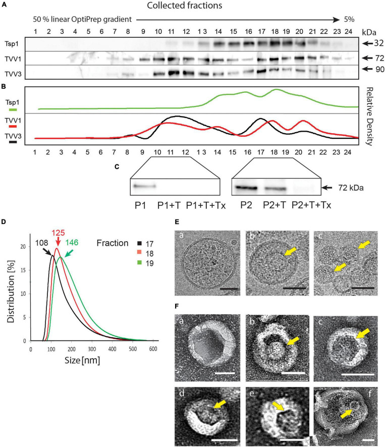
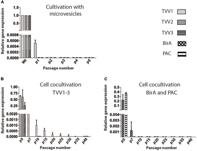
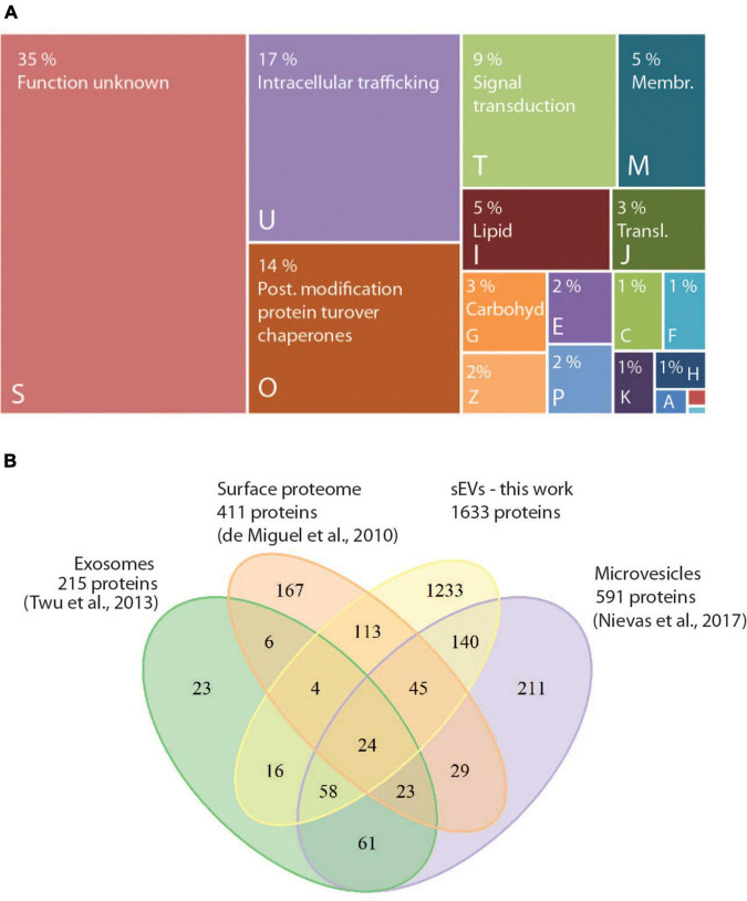
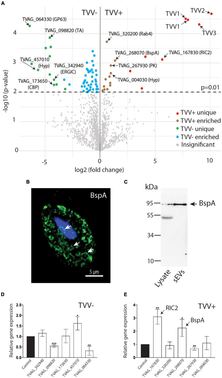
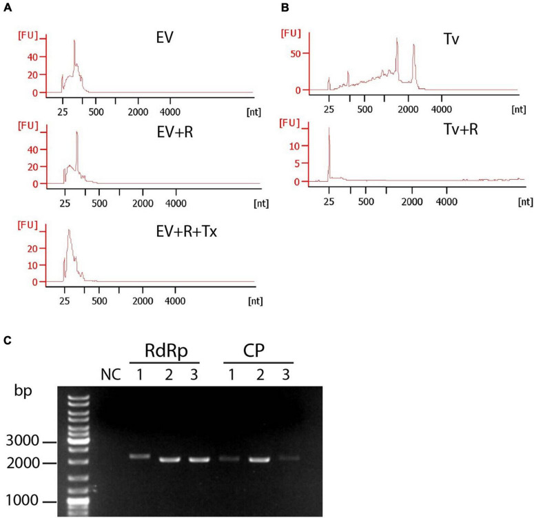
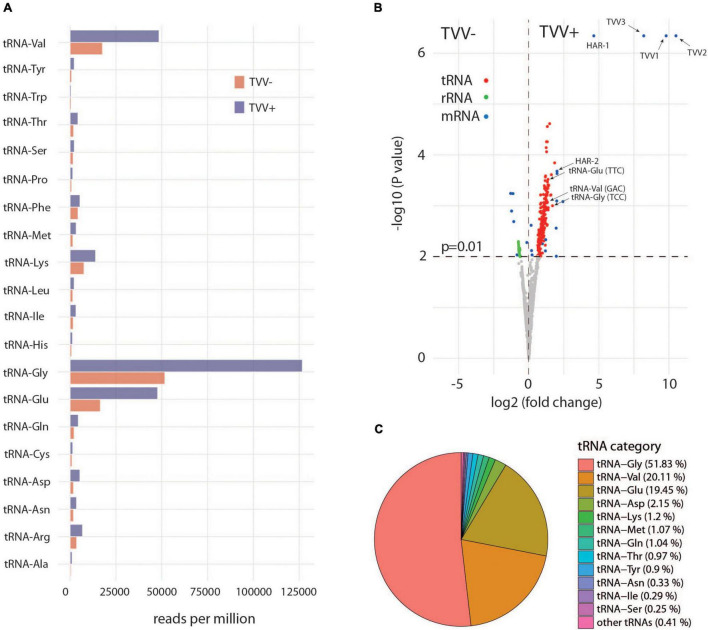
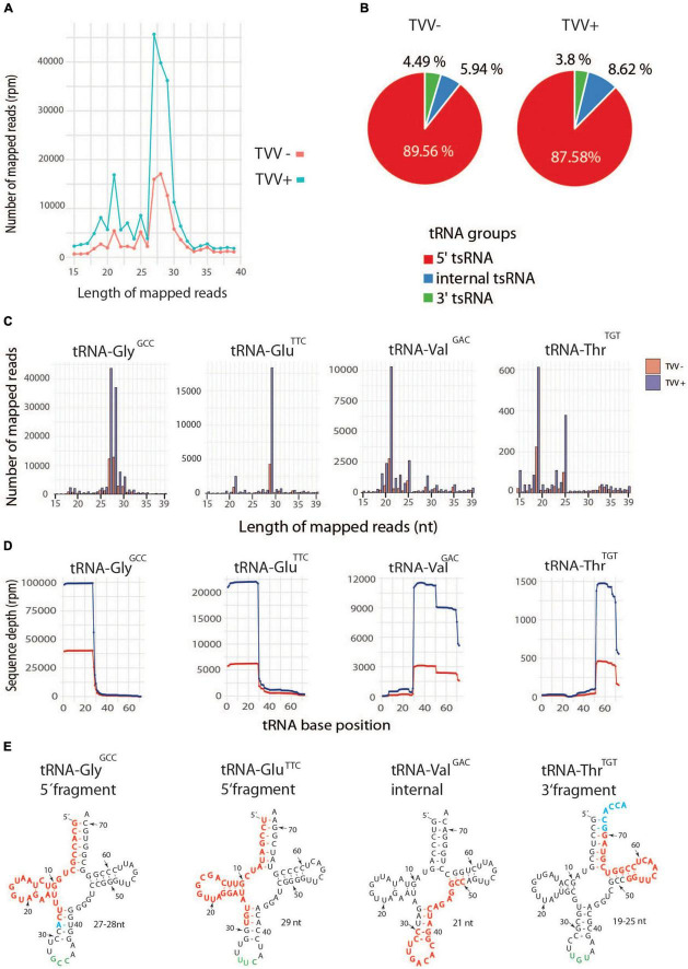
Trichomonas vaginalis is a parasitic protist that infects the human urogenital tract. During the infection, trichomonads adhere to the host mucosa, acquire nutrients from the vaginal/prostate environment, and release small extracellular vesicles (sEVs) that contribute to the trichomonad adherence and modulate the host-parasite communication. Approximately 40-70% of T. vaginalis strains harbor a double-stranded RNA virus called Trichomonasvirus (TVV). Naked TVV particles have the potential to stimulate a proinflammatory response in human cells, however, the mode of TVV release from trichomonads to the environment is not clear. In this report, we showed for the first time that TVV particles are released from T. vaginalis cells within sEVs. The sEVs loaded with TVV stimulated a higher proinflammatory response of human HaCaT cells in comparison to sEVs from TVV negative parasites. Moreover, a comparison of T. vaginalis isogenic TVV plus and TVV minus clones revealed a significant impact of TVV infection on the sEV proteome and RNA cargo. Small EVs from TVV positive trichomonads contained 12 enriched and 8 unique proteins including membrane-associated BspA adhesine, and about a 2.5-fold increase in the content of small regulatory tsRNA. As T. vaginalis isolates are frequently infected with TVV, the release of TVV via sEVs to the environment represents an important factor with the potential to enhance inflammation-related pathogenesis during trichomoniasis.

Keywords: TVV; Trichomonasvirus; exosome; extracellular vesicle; proteomics; tsRNA.


Conflict of interest statement

The authors declare that the research was conducted in the absence of any commercial or financial relationships that could be construed as a potential conflict of interest.

Figures

FIGURE 1
FIGURE 1
Small EVs produced by Trichomonas vaginalis TV79-49c1+ contain Trichomonasvirus (TVV) particles. (A) Immunoblot analysis of 24 fractions collected from the last step of EV separation using linear OptiPrep gradient. Antibodies against Tsp1 (sEV marker), and TVV1 and TVV3 capsid proteins were used for screening of fractions. (B) Densitometry of western blots. (C) Protein protection assay. Membrane protection of TVV proteins was monitored by immunoblotting using antibodies against TVV1 capsid protein. P1, the pool of fractions 10–12; P2, the pool of fractions 17–19; T, trypsin; Tx, Triton X100. (D) Multi-angle dynamic light scattering analysis of sEV size in fractions 17, 18, and 19. (E) (a–c) Cryo-electron microscopy of sEV fraction. (F) (a–f). Electron microscopy of sEVs after negative staining. Arrows point to TVV particles. Bars indicate 40 nm.
FIGURE 2
FIGURE 2
TVV transmission experiments. (A) Monitoring of TVVs by RT PCR in TV79-49c1 clone after incubation with sEVs from TV79-49c1+. (B) RT PCR monitoring of TVVs, in TV79-49c1 acceptor clone for 40 subcultures after co-cultivation with TVV79-49c1+ donor clone. TVV- plus clone was transformed with a plasmid for expression of puromycin N-acetyl transferase (PAC), and biotin ligase (BirA), and the TVV-minus clone was resistant to geneticin. The clones were mixed, co-cultivated for five subcultures, and then geneticin was added for 35 subcultures. (C) RT PCR monitoring of genes for BirA and PAC.
FIGURE 3
FIGURE 3
Assignment of protein in the proteome of sEVs to functional categories. (A) COG (Clusters of orthologous groups) functional categories of identified proteins in sEVs from T. vaginalis TV79-49c1+ and TV79-49c1. Capital letters correspond to COG categories: A, RNA processing and modification; B, Chromatin structure and dynamics; C, Energy production and conversion; E, Amino acid transport and metabolism; F, Nucleotide transport and metabolism; G, Carbohydrate transport and metabolism; H, Coenzyme transport and metabolism; I, Lipid transport and metabolism; -J, Translation, ribosomal structure and biogenesis; K, Transcription; M, Cell wall, membrane, envelope biogenesis; O, Posttranslational modification, protein turnover, chaperones; P, Inorganic ion transport and metabolism; S, unknown function; T, Signal transduction mechanism; U, Intracellular trafficking, secretion, and vesicular transport; Z, Cytoskeleton. (B) Venn diagram comparing proteomes of sEVs, exosomes (Twu et al., 2013), surface proteome (De Miguel et al., 2010), and microvesicles (Nievas et al., 2018).
FIGURE 4
FIGURE 4
Comparison of sEVs proteomes from TV79-49c1 (TVV–) and TV79-49c1+(TVV +). (A) Volcano plot analysis of proteomes using statistical significance p-value 0.01. (B) Structured illumination microscopy of T. vaginalis TV79-49c1+ expressing HA-tagged BspA (TVAG_268070). Arrows indicate large vesicles reminiscent of multivesicular bodies. (C) Immunoblot detection of HA-tagged BspA (TVAG_268070) in cell lysate and sEVs from TV79-49c1+ transformed T. vaginalis. BspA corresponds to 87 kDa protein in the cell lysate and sEVs. An additional band of 50 kDa in the lysate is likely a product of BspA cleavage. D, E. RT PCR of selected genes for proteins, which were unique or upregulated in proteomes of TVV– (D) or TVV+ (E) sEVs. CBP, calcium-binding protein; ERGIC, endoplasmic reticulum-Golgi intermediate compartment protein; GP63, leishmanolysin-like metallopeptidase; hyp, conserved hypothetical protein; PK, CMGC family protein kinase; RIC2, repair of iron centers protein 2; TA, tyrosine aminotransferase. *p-value < 0.05, **p-value < 0.005, ***p-value = 0.0005.
FIGURE 5
FIGURE 5
Protection assay of sRNAs in sEVs of TV79-49c1+. (A) Small RNAs were treated with RNAse A (EV + R) or RNAse A and Triton X-100 (EV + R + Tx). Arrows point to a prominent peak of 100 nt. (B) Treatment of T. vaginalis cellular RNAs (Tv) with RNAse A (Tv + R). RNA was analyzed using Bioanalyzer. (C) Amplification of ORFs for RdRp and capsid protein (CP) of TVV1, TVV2, and TVV3 from RNA isolated from sEVs.
FIGURE 6
FIGURE 6
Transfer RNAs are enriched in TV79-49c1+. (A) Distribution of tRNA types in sEVs from TV79-49c1 (TVV-), and TV79-49c1+ (TVV +). (B) Volcano plot analysis of tRNAs, rRNAs, and mRNAs in TVV- and TVV+ sEVs. (C) Distribution of tRNA types that were significantly enriched in TVV+ sEVs in comparison to TVV- sEVs.
FIGURE 7
FIGURE 7
Enrichment of tsRNA in TVV + sEVs. (A) Size distribution of total tRNA fragments in TVV-, and TVV+ sEVs. (B) Distribution of fragment categories in TVV + sEVs. (C) Size distribution of the most frequent tsRNAs in TVV- and TVV+ sEVs. (D) Coverage of dominant tsRNAs that were mapped to corresponding tRNAs. Transfer sRNAs were grouped based on tRNA type and anticodon. (E) Mapping of most frequent tsRNA fragments to mature tRNAs. Red letters indicate a sequence of tsRNA, blue letters indicate extension in some sequences.
FIGURE 8
FIGURE 8
Small EVs from TV79-49c1+ stimulate a proinflammatory response in HaCaT cells. HaCaT cells were incubated with sEVs from TV79-49c1 or Tv79-49c1+ for 24 h and subsequently levels of immune mediators were determined in a conditioned medium using ELISA. Medium without sEVs was used as a negative control. ***p-value < 0.0001.

Similar articles

Cited by

References

    1. Abramowicz A., Story M. D. (2020). The long and short of it: the emerging roles of non-coding RNA in small extracellular vesicles. Cancers 12:1445. 10.3390/cancers12061445 - DOI - PMC - PubMed
    1. Ahsan N. A., Sampey G. C., Lepene B., Akpamagbo Y., Barclay R. A., Iordanskiy S., et al. (2016). Presence of viral RNA and proteins in exosomes from cellular clones resistant to Rift Valley fever virus infection. Front. Microbiol. 7:139. 10.3389/fmicb.2016.00139 - DOI - PMC - PubMed
    1. Alderete J. F. (1999). Iron modulates phenotypic variation and phosphorylation of P270 in double-stranded RNA virus-infected Trichomonas vaginalis. Infect. Immun. 67 4298–4302. 10.1128/IAI.67.8.4298-4302.1999 - DOI - PMC - PubMed
    1. Amos B., Aurrecoechea C., Barba M., Barreto A., Basenko E. Y., Bażant W., et al. (2022). VEuPathDB: the eukaryotic pathogen, vector and host bioinformatics resource center. Nucleic Acids Res. 50 D898–D911. 10.1093/nar/gkab929 - DOI - PMC - PubMed
    1. Artuyants A., Campos T. L., Rai A. K., Johnson P. J., Dauros-Singorenko P., Phillips A., et al. (2020). Extracellular vesicles produced by the protozoan parasite Trichomonas vaginalis contain a preferential cargo of tRNA-derived small RNAs. Int. J. Parasitol. 50 1145–1155. 10.1016/J.IJPARA.2020.07.003 - DOI - PubMed