. 2021 Dec 8:1:58.
doi: 10.1038/s43856-021-00059-3. eCollection 2021.

Specific ablation of PDGFRβ-overexpressing pericytes with antibody-drug conjugate potently inhibits pathologic ocular neovascularization in mouse models

Affiliations

Specific ablation of PDGFRβ-overexpressing pericytes with antibody-drug conjugate potently inhibits pathologic ocular neovascularization in mouse models

Seok Jae Lee et al. Commun Med (Lond). .

Abstract

Background: Crosstalk between pericytes and endothelial cells is critical for ocular neovascularization. Endothelial cells secrete platelet-derived growth factor (PDGF)-BB and recruit PDGF receptor β (PDGFRβ)-overexpressing pericytes, which in turn cover and stabilize neovessels, independent of vascular endothelial growth factor (VEGF). Therapeutic agents inhibiting PDGF-BB/PDGFRβ signaling were tested in clinical trials but failed to provide additional benefits over anti-VEGF agents. We tested whether an antibody-drug conjugate (ADC) - an engineered monoclonal antibody linked to a cytotoxic agent - could selectively ablate pericytes and suppress retinal and choroidal neovascularization.

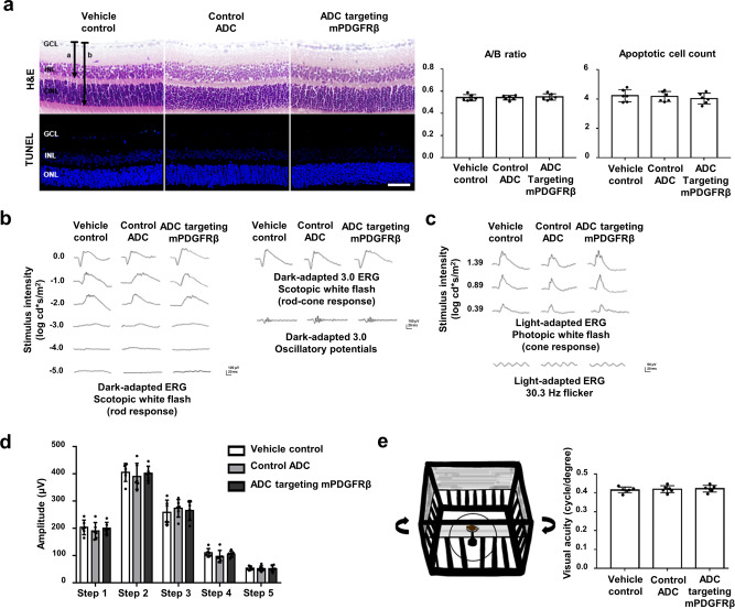
Methods: Immunoblotting, flow cytometry, cell viability test, and confocal microscopy were conducted to assess the internalization and cytotoxic effect of ADC targeting mPDGFRβ in an in vitro setting. Immunofluorescence staining of whole-mount retinas and retinal pigment epithelium-choroid-scleral complexes, electroretinography, and OptoMotry test were used to evaluate the effect and safety of ADC targeting mPDGFRβ in the mouse models of pathologic ocular neovascularization.

Results: ADC targeting mPDGFRβ is effectively internalized into mouse brain vascular pericytes and showed significant cytotoxicity compared with the control ADC. We also show that specific ablation of PDGFRβ-overexpressing pericytes using an ADC potently inhibits pathologic ocular neovascularization in mouse models of oxygen-induced retinopathy and laser-induced choroidal neovascularization, while not provoking generalized retinal toxicity.

Conclusion: Our results suggest that removing PDGFRβ-expressing pericytes by an ADC targeting PDGFRβ could be a potential therapeutic strategy for pathologic ocular neovascularization.

Keywords: Drug delivery; Target validation.


Conflict of interest statement

Competing interestsThe authors declare no competing interests.

Figures

Fig. 1
Fig. 1. PDGFRβ is highly expressed on pericytes in pathologic neovessels of OIR and LI-CNV.
Representative immunofluorescence images of the retinal vasculature in a wild-type (control) mouse (a) and an oxygen-induced retinopathy (OIR) mouse (b). Immunofluorescence images of anti-isolectin B4 (IB4, red), anti-neural/glial antigen 2 (NG2, green), platelet-derived growth factor receptor β (PDGFRβ, gray), and 4′,6-diamidino-2-phenylindole (DAPI, blue) showing that PDGFRβ expression is mainly localized in the retinal vessels and surrounding tissue. In the OIR mouse, PDGFRβ is significantly overexpressed in the vascular tufts (b). Representative immunofluorescence images of retinal pigment epithelium (RPE) in a wild-type (control) mouse (c) and a mouse with laser-induced choroidal neovascularization (LI-CNV) (d). The choroidal neovascularization (CNV) lesion in the LI-CNV mouse shows strong PDGFRβ expression (d). Scale bar, 200 μm.
Fig. 2
Fig. 2. Preparation and characterization of ADC targeting mPDGFRβ.
a Flow cytometry of bispecific anti-mouse platelet-derived growth factor receptor β (mPDGFRβ) × cotinine single-chain variable fragment (scFv)- kappa constant (Cκ)-scFv fusion proteins (anti-mPDGFRβ × cotinine). Mouse brain vascular pericytes (MBVPs) were treated with anti-mPDGFRβ × cotinine and probed with APC-conjugated anti-human Cκ antibody. As a control, anti-human epidermal growth factor receptor 2 (HER2) × cotinine scFv-Cκ-scFv fusion protein (anti-HER2 × cotinine) was used; anti-HER scFv was derived from trastuzumab. b Confocal microscopy of the internalization of anti-mPDGFRβ × cotinine. MBVP cells were incubated with scFv-Cκ-scFv fusion proteins with or without mPDGF-BB. Scale bars in the inset image and on the bottom right correspond to 1 μm and 10 μm, respectively. c Cytotoxicity of antibody-drug conjugate (ADC) targeting mPDGFRβ. MBVP cells were treated with ADC in the absence (−) or presence (+) of mPDGF-BB. The relative cell viability was calculated using the cellular ATP level. Anti-HER2 × cotinine complexed with cotinine-duocarmycin was used as a control ADC and drug only refers to cotinine-duocarmycin in fresh media. The results are shown as means ± standard deviation (SD) from triplicate experiments.
Fig. 3
Fig. 3. ADC targeting mPDGFRβ ameliorates retinal neovascularization in the OIR model.
a Schematic experimental timeline of antibody-drug conjugate (ADC) administration and tissue preparation in the oxygen-induced retinopathy (OIR) mouse model. OIR-induced pups were treated with vehicle control, control ADC, or ADC targeting mouse platelet-derived growth factor receptor β (mPDGFRβ) through intravitreal injection at P14. Three days after treatment, the mouse eyeballs were dissected to prepare whole-mounted retina samples. Phosphate-buffered saline (PBS) was used as the vehicle control. Anti-human epidermal growth factor receptor 2 (HER2) × cotinine complexed with cotinine-duocarmycin was used as a control ADC; anti-HER2 scFv was derived from trastuzumab. b Representative immunofluorescence images of whole-mounted retina samples from OIR pups stained with isolectin B4 (IB4, red) and neural/glial antigen 2 (NG2, green) to visualize the retinal vessels and pericytes. Scale bars on the top, middle, and bottom correspond to 500 μm, 100 μm, and 150 μm, respectively. c Quantification of the avascular area and neovascular tuft area. The avascular area and neovascular tuft area were quantified and presented as percentages of the total area of the retina. All data were analyzed using NIH ImageJ software, and values are presented as percentages of the mean ± SEM (n = 6 mice for each data set). *P < 0.01, **P < 0.001, obtained using one-way analysis of variance (ANOVA) and Tukey’s post-hoc tests. d Quantification of NG2 coverage of IB4 + vessels at the mid-peripheral region (pathological neovascular tuft area, left) and peripheral region (peripheral vascularized area, right), respectively in the whole-mounted retinas. All data were analyzed using a built-in measuring tool, the LAS X system (Leica Microsystems Ltd., Wetzlar, Germany). Error bars represent standard error mean (SEM, n = 6 mice for each data set). **P < 0.001, obtained using one-way ANOVA and Tukey’s post-hoc tests. Source data are provided as a Supplementary Data file.
Fig. 4
Fig. 4. ADC targeting mPDGFRβ inhibits laser-induced CNV.
a Schematic experimental timeline of ADC administration and tissue preparation in the laser induced-choroidal neovascularization (LI-CNV) mouse model. Six-week-old wild-type C57BL/6J male mice received laser photocoagulation and were treated with vehicle control, control antibody-drug conjugate (ADC), or ADC targeting mouse platelet-derived growth factor receptor β (mPDGFRβ) through intravitreal injection at 7 days. Three days after treatment, the mouse eyeballs were dissected, and RPE-choroid-scleral (RCS) complexes were whole mounted. Phosphate-buffered saline (PBS) was used as the vehicle control. Anti-human epidermal growth factor receptor-2 (HER2) × cotinine complexed with cotinine-duocarmycin was used as a control ADC; anti-HER2 scFv was derived from trastuzumab. b Representative images of CNV at 10 days after laser photocoagulation and immunostaining with isolectin B4 (IB4), neural/glial antigen 2 (NG2), and PDGFRβ. Scale bar, 200 μm. c Quantitation of CNV volume, CNV area, NG2 volume, and PDGFRβ volume. All quantitative data were measured using the built-in tools, the LAS X systems (Leica Microsystems, Wetzlar, Germany). Each value represents the mean ± standard error mean (SEM, n = 6 mice for each group set). *P < 0.01, obtained using one-way analysis of variance (ANOVA) and Tukey’s post-hoc tests. Source data are provided as a Supplementary Data file.
Fig. 5
Fig. 5. Retinal safety evaluation in mice receiving ADC targeting mPDGFRβ.
a Representative hematoxylin and eosin (H&E) and terminal deoxynucleotidyl transferase dUTP nick end labeling (TUNEL) staining images of retinal tissues at 7 days after injection of vehicle control, control antibody-drug conjugate (ADC), or ADC targeting mouse platelet-derived growth factor receptor β (mPDGFRβ). Scale bar, 500 μm. Phosphate-buffered saline (PBS) was used as the vehicle control. Anti-human epidermal growth factor receptor 2 (HER2) × cotinine complexed with cotinine-duocarmycin was used as a control ADC; anti-HER2 single-chain variable fragment (scFv) was derived from trastuzumab. Each value represents the mean ± standard error mean (SEM, n = 6 mice per data set). b The overall stimulus parameters used in the study for the representative group of averaged dark-adapted electroretinography (ERG) waveforms were as follows: step 1: scotopic white flash; step 2: scotopic white flash 3.0 cd·s/m2; and step 3: oscillatory potential, 3.0 cd·s/m2. c The overall stimulus parameters used in the study for the representative group-averaged light-adapted ERG waveforms were as follows: step 4: photopic white flash; and step 5: 30.3 Hz flicker. d Amplitudes of the b-wave by steps at 7 days after the injection of PBS, control ADC, or ADC targeting mPDGFRβ. Anti-HER2 × cotinine complexed with cotinine-duocarmycin was used as a control ADC; anti-HER2 scFv was derived from trastuzumab. Each value represents the mean ± standard error mean (SEM, n = 6 mice per data set). e Schematic diagram depicting the OptoMotry test and spatial frequency thresholds, in cycles per degree. Each value represents the mean ± SEM (n = 6 mice per data set). No significant differences were found in the retinal layer thickness ratio, terminal deoxynucleotidyl transferase dUTP nick end labeling (TUNEL) assay, ERG amplitudes, or spatial frequency of the OptoMotry test among the three groups, as confirmed by one-way analysis of variance (ANOVA) and Tukey’s post-hoc tests. Source data are provided as a Supplementary Data file.

References

    1. Ferrara N. Vascular endothelial growth factor and age-related macular degeneration: from basic science to therapy. Nat. Med. 2010;16:1107–1111. doi: 10.1038/nm1010-1107. - DOI - PubMed
    1. Ferrara N, Adamis AP. Ten years of anti-vascular endothelial growth factor therapy. Nat. Rev. Drug Discov. 2016;15:385–403. doi: 10.1038/nrd.2015.17. - DOI - PubMed
    1. Ohno-Matsui K, Ikuno Y, Lai TYY, Gemmy Cheung CM. Diagnosis and treatment guideline for myopic choroidal neovascularization due to pathologic myopia. Prog. Retin. Eye Res. 2018;63:92–106. doi: 10.1016/j.preteyeres.2017.10.005. - DOI - PubMed
    1. Yang J, Yuan M, Xia S, Chen Y. Six-year real-world outcomes of antivascular endothelial growth factor monotherapy and combination therapy for various subtypes of polypoidal choroidal vasculopathy. J. Ophthalmol. 2019;2019:1609717. doi: 10.1155/2019/1609717. - DOI - PMC - PubMed
    1. Aiello LP, et al. Vascular endothelial growth factor in ocular fluid of patients with diabetic retinopathy and other retinal disorders. New Engl. J. Med. 1994;331:1480–1487. doi: 10.1056/NEJM199412013312203. - DOI - PubMed