. 2021 Nov 15:1:47.
doi: 10.1038/s43856-021-00047-7. eCollection 2021.

A case report describing the immune response of an infant with congenital heart disease and severe COVID-19

Affiliations

A case report describing the immune response of an infant with congenital heart disease and severe COVID-19

Danielle Wurzel et al. Commun Med (Lond). .

Abstract

Background: Children with SARS-CoV-2 infection generally present with milder symptoms or are asymptomatic in comparison with adults, however severe disease occurs in a subset of children. To date, the immune correlates of severe COVID-19 in young children have been poorly characterised.

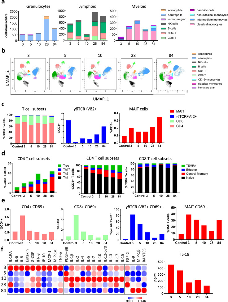
Methods: We report the kinetics of immune responses in relation to clinical and virological features in an infant with acute severe COVID-19 using high-dimensional flow cytometry and multiplex cytokine analysis.

Results: Systemic cellular and cytokine profiling show an initial increase in neutrophils and monocytes with depletion of lymphoid cell populations (particularly CD8 + T and NK cells) and elevated inflammatory cytokines. Expansion of memory CD4 + T (but not CD8 + T) cells occurred over time, with a predominant Th2 bias. Marked activation of T cell populations observed during the acute infection gradually resolved as the child recovered. Substantial in vitro activation of T-cell populations and robust cytokine production, in response to inactivated SARS-CoV-2 stimulation, was observed 3 months after infection indicating durable, long-lived cellular immune memory.

Conclusions: These findings provide important insights into the immune response of a young infant with severe COVID-19 and will help to inform future research into therapeutic targets for high-risk groups.

Keywords: Paediatric research; Viral infection.


Conflict of interest statement

Competing interestsThe authors declare no competing interests.

Figures

Fig. 1
Fig. 1. Clinical and laboratory characteristics of an infant with severe COVID-19.
A Clinical timeline showing immuno-modulatory therapies in relation to clinical course. B Plain chest film performed on admission illustrating bilateral interstitial infiltrates with left lower lobe collapse and consolidation. C Virologic findings illustrating SARS-CoV-2 Cycle threshold Ct values in naso-/oropharyngeal and urine specimens that reduce in association with clinical improvement. D Serum IgG, IgM, and IgA to S1 using in-house ELISA modified from Mount Sinai Laboratories, USA; and microneutralisation assay demonstrating a rapid rise in neutralising antibody titres. E Elevated ferritin and LDH with initial hyper-inflammatory picture and rapid reduction in ferritin coinciding with clinical improvement. F Persistent lymphopaenia; initial high IL-6 with reduction after administration of tocilizumab. IVIG intravenous immunoglobulin, LDH lactate dehydrogenase, NPA nasopharyngeal aspirate, EU ELISA units, Ct cycle threshold.
Fig. 2
Fig. 2. Immune cell profiling in whole blood and PBMCs.
A Flow cytometry was performed on whole blood samples collected at 3, 5, 10, 28, and 84 days following admission. Neutrophils, eosinophils, CD4 T cells, CD8 T cells, B cells, Natural Killer (NK) cells, monocytes (classical, intermediate, and non-classical), and dendritic cells were classified by manual gating and expressed as cells/µL using counting beads. B Unsupervised clustering and dimensionality reduction were performed using FlowSOM and UMAP on a concatenated file containing 150,000 live single cells (30,000 randomly selected cells from each time point). The UMAP plots at each time point are coloured according to the generated FlowSOM clusters. C Flow cytometry was performed on peripheral blood mononuclear cells collected on days 3, 5, 10, 28, and 84 following admission. CD4, CD8, γδVδ2+ T-cells, and MAIT cells were classified by manual gating and expressed as a frequency of CD3+ T-cells. These results were compared to an age/sex-matched control, obtained at a single time-point. D Subsets of CD4+ and CD8+ T-cells were further categorised by flow cytometry to determine the frequency of Th1, Th2, Th17, Treg, and memory subsets. These were expressed as the frequency of CD4+ or CD8+ T-cells. E Flow cytometry was performed to assess the activation status of T-cells. CD69+ was expressed as a proportion of total CD4+, CD8+, γδVδ2+ T-cells, and MAIT cells. F Cytokines were quantified in the serum using a multiplex cytokine assay and visualised in a heatmap containing log2 transformed values and clustered according to peak expression at day 3 (cluster 1), day 5 (cluster 2), day 10 (cluster 3), and day 28 (cluster 4). IL-18 levels at days 3, 5, 10, 28, and 84 were quantified by ELISA and expressed as pg/ml. UMAP Uniform Manifold Approximation and Projection, MAIT mucosal-associated invariant T cells.
Fig. 3
Fig. 3. Immune response to inactivated SARS-CoV-2.
A Flow cytometry was performed on PBMCs following 4-day stimulation with inactivated SARS-CoV-2 to observe T-cell activation status. CD69+ was expressed as a proportion of total CD4+ or CD8+ T-cells. B Cytokines were quantified in PBMC supernatants following 4-day stimulation with inactivated SARS-CoV-2 by multiplex cytokine assay to assess memory T-cell responses.

References

    1. Leidman, E. et al. COVID-19 Trends among persons aged 0–24 years—United States, March 1–December 12, 2020. MMWR Morb. Mortal Wkly Rep.70, 88−94 (2021). - PMC - PubMed
    1. Cowley D, Donato CM, Roczo-Farkas S, Kirkwood CD. Novel G10P[14] rotavirus strain, northern territory, Australia. Emerg. Infect. Dis. 2013;19:1324–1327. doi: 10.3201/eid1908.121653. - DOI - PMC - PubMed
    1. Chan, J. F. et al. Improved molecular diagnosis of COVID-19 by the novel, highly sensitive, and specific COVID-19-RdRp/Hel real-time reverse transcription-PCR assay validated in vitro and with clinical specimens. J. Clin. Microbiol.58, e00310−20 (2020). - PMC - PubMed
    1. Corman, V. M., et al. Detection of 2019 novel coronavirus (2019-nCoV) by real-time RT-PCR. Euro Surveill25, 2000045 (2020). - PMC - PubMed
    1. Tay MZ, Poh CM, Rénia L, MacAry PA, Ng LFP. The trinity of COVID-19: immunity, inflammation, and intervention. Nat. Rev. Immunol. 2020;20:363–374. doi: 10.1038/s41577-020-0311-8. - DOI - PMC - PubMed