. 2022 Apr 28;25(5):104314.
doi: 10.1016/j.isci.2022.104314. eCollection 2022 May 20.

CDX2-induced intestinal metaplasia in human gastric organoids derived from induced pluripotent stem cells

Affiliations

CDX2-induced intestinal metaplasia in human gastric organoids derived from induced pluripotent stem cells

Takahiro Koide et al. iScience. .

Abstract

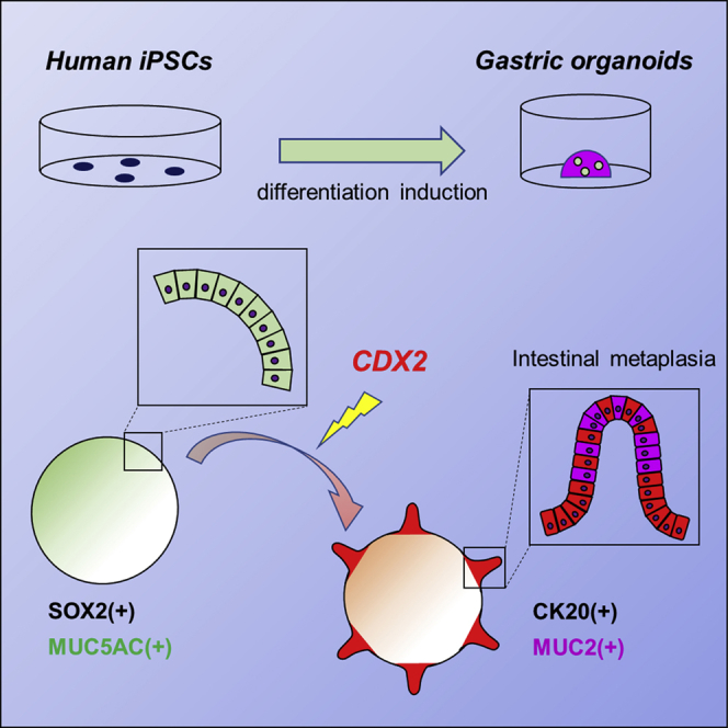
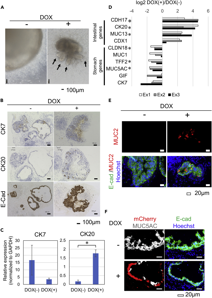
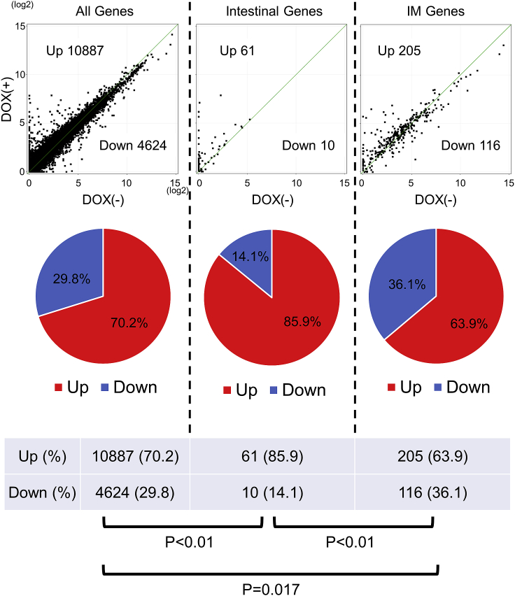
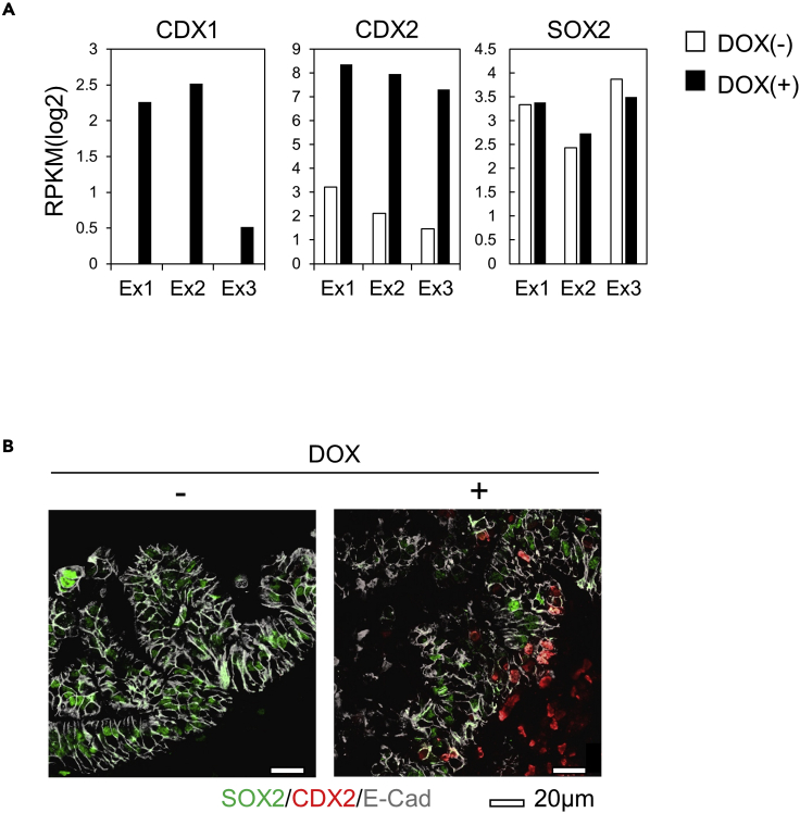
Intestinal metaplasia is related to gastric carcinogenesis. Previous studies have suggested the important role of CDX2 in intestinal metaplasia, and several reports have shown that the overexpression of CDX2 in mouse gastric mucosa caused intestinal metaplasia. However, no study has examined the induction of intestinal metaplasia using human gastric mucosa. In the present study, to produce an intestinal metaplasia model in human gastric mucosa in vitro, we differentiated human-induced pluripotent stem cells (hiPSC) to gastric organoids, followed by the overexpression of CDX2 using a tet-on system. The overexpression of CDX2 induced, although not completely, intestinal phenotypes and the enhanced expression of many, but not all, intestinal genes and previously reported intestinal metaplasia-related genes in the gastric organoids. This model can help clarify the mechanisms underlying intestinal metaplasia and carcinogenesis in human gastric mucosa and develop therapies to restitute precursor conditions of gastric cancer to normal mucosa.

Keywords: Biological sciences; Cell biology; Stem cells research.


Conflict of interest statement

The authors declare no competing interests.

Figures

None
Graphical abstract
Figure 1
Figure 1
The production of the PB-TAC-CDX2-ERN vector and introduction into iPSC (A) A schematic diagram of the piggyBac vector containing doxycycline (DOX)-inducible CDX2 (PB-TAC-CDX2-ERN). (B) The cell morphologies of CDX2-iPSC (left panels) and iPSC before transfection of the PB-TAC-CDX2-ERN vector: Parental-iPSC (right panels). Scale bars: black bars = 500 μm, white bars = 50 μm. (C) An RT-PCR analysis showed that CDX2-iPSC maintained the mRNA expression of the pluripotent markers OCT3/4, SOX2, and NANOG. GAPDH was used as an endogenous control. RT: reverse transcriptase. (D) Immunostaining showed that CDX2-iPSC (upper panels) as well as Parental-iPSC (lower panels) expressed the pluripotency markers NANOG, OCT3/4, and SOX2. The nuclei were stained blue with Hoechst33342. Scale bars, 50 μm.
Figure 2
Figure 2
DOX-inducible CDX2 in CDX2-iPSC (A) A fluorescence analysis of the mCherry expression in CDX2-iPSC with (lower panels) or without (upper panels) DOX administration for 24 h. The representative data of three independent experiments are shown. Scale bars: black bars = 20 μm, white bars = 200 μm. (B) An RT-PCR analysis of the total CDX2 expression in CDX2-iPSC. The colorectal cancer cell line Colo320 was used as a positive control of the CDX2 expression. GAPDH was used as an endogenous control. CDX2-iPSC in culture with DOX administration showed CDX2 expression. Representative data of two independent experiments are shown. (C) Western blotting showing the protein expressions of CDX2 and β-actin in CDX2-iPSC cultured with or without DOX administration as well as Colo320. Cell lysates were collected at 2 days post-tet-on. Representative data of three independent experiments are shown. (D) Immunostaining of CDX2 in CDX2-iPSC at 2 days post-DOX administration. CDX2 was expressed in CDX2-iPSC cultured with DOX and co-localized with mCherry. Scale bars, 200 μm.
Figure 3
Figure 3
The overexpression of CDX2 in induced gastric organoids (A) A schematic representation of the protocol of gastric organoid differentiation from CDX2-iPSC. The administration of DOX (1 μM) started between days 35 and 40. (B) Immunofluorescence analyses of the MUC5AC and E-cadherin (E-Cad) expression in differentiated gastric organoids at day 37 without DOX treatment. Scale bar, 40 μm. (C) Morphologies of gastric organoids derived from CDX2-iPSC after 1 μM DOX treatment for 10 days from Day 36. The representative data of three independent experiments are shown. Scale bars, 200 μm. (D) An RT-PCR analysis showed the mRNA expression of CDX2 at Day45 with or without DOX treatment for 9 days. The expression of CDX2 was increased in DOX(+) organoids. GAPDH was used as an endogenous control. (E) Immunofluorescence analyses of CDX2 and mCherry in gastric organoids derived from CDX2-iPSC at Day45 with or without DOX treatment for 10 days. Scale bars, 20 μm.
Figure 4
Figure 4
Phenotype alterations of DOX(+) organoids (A) Phase contrast micrographs of the morphologies of DOX(−) organoids (Day 42, left panel) and DOX(+) organoids (Day 42, right panel). Arrows indicate crypt-like structures. Scale bars, 100 μm. (B) Immunohistochemical analyses of CK7, CK20, and E-Cadherin in the gastric organoids derived from CDX2-iPSC at day 40 with (+) or without (−) DOX treatment for 5 days. Scale bars, 100 μm. (C) qRT-PCR analysis showed the mRNA expressions of CK7 and CK20. The expression of CK20 was increased in DOX(+) organoids. The mRNA expression was normalized to GAPDH. Data are represented as mean ± SEM of three independent induction experiments. ∗, p < 0.05. (D) The difference in the expression of the intestinal markers and the stomach markers between DOX(+) and DOX(−) organoids was calculated by subtracting the log2 RPKM value of DOX(−) from that of DOX(+). ∗, p < 0.05. (E) Immunofluorescence analyses of E-Cadherin and MUC2 expression in the gastric organoids derived from CDX2-iPSC at Day 40 with (+) or without (−) DOX treatment for 5 days. Scale bars, 20 μm. (F) Immunofluorescence analyses of E-Cadherin and MUC5AC in gastric organoids from CDX2-iPSC at Day 45 with (+) (lower panels) or without (−) (upper panels) DOX treatment for 10 days. Scale bars, 20 μm.
Figure 5
Figure 5
Analyses of the global gene expression changes in CDX2-overexpressing gastric organoids Scatter plot analyses of the all genes (left panel), highly expressed genes in the normal intestine compared to the normal stomach (“Intestinal Genes,” middle panel) and gene for intestinal metaplasia marker genes (Lee et al., 2010) (“IM Genes,” right panel) in DOX(+) gastric organoids (y axis, n = 3) versus DOX(−) gastric organoids (x axis, n = 3) after the addition of DOX for 7 days. Pie charts (middle panels) and tables (lower panels) show the percentage of genes that showed an increased or a decreased expression.
Figure 6
Figure 6
The expression of the master transcription factors CDX1, CDX2, and SOX2 (A) RPKM values (log2) of CDX1, CDX2, and SOX2 in RNA-seq data. (B) Immunofluorescence analyses of E-Cadherin, SOX2, and CDX2 in gastric organoids from CDX2-iPSC at Day 40 without (−) (left panel) or with (+) (right panel) DOX treatment for 5 days, using confocal microscopy. Scale bars, 20 μm.

Similar articles

Cited by

References

    1. Almeida R., Silva E., Santos-Silva F., Silberg D.G., Wang J., De Bolos C., David L. Expression of intestine-specific transcription factors, CDX1 and CDX2, in intestinal metaplasia and gastric carcinomas. J. Pathol. 2003;199:36–40. doi: 10.1002/path.1246. - DOI - PubMed
    1. Aoi T., Yae K., Nakagawa M., Ichisaka T., Okita K., Takahashi K., Chiba T., Yamanaka S. Generation of pluripotent stem cells from adult mouse liver and stomach cells. Science. 2008;321:699–702. doi: 10.1126/science.1154884. - DOI - PubMed
    1. Barker N., Huch M., Kujala P., van de Wetering M., Snippert H.J., van Es J.H., Sato T., Stange D.E., Begthel H., van den Born M., et al. Lgr5(+ve) stem cells drive self-renewal in the stomach and build long-lived gastric units in vitro. Cell Stem Cell. 2010;6:25–36. doi: 10.1016/j.stem.2009.11.013. - DOI - PubMed
    1. Beck F., Chawengsaksophak K., Waring P., Playford R.J., Furness J.B. Reprogramming of intestinal differentiation and intercalary regeneration in Cdx2 mutant mice. Proc. Natl. Acad. Sci. U S A. 1999;96:7318–7323. doi: 10.1073/pnas.96.13.7318. - DOI - PMC - PubMed
    1. Chen H.Y., Hu Y., Lu N.H., Zhu Y. Caudal type homeoboxes as a driving force in Helicobacter pylori infection-induced gastric intestinal metaplasia. Gut Microbes. 2020;12:1809331–1809412. doi: 10.1080/19490976.2020.1809331. - DOI - PMC - PubMed