

Current methods for fabricating 3D cardiac engineered constructs
- PMID: 35602954
- PMCID: PMC9118671
- DOI: 10.1016/j.isci.2022.104330
Current methods for fabricating 3D cardiac engineered constructs
Abstract
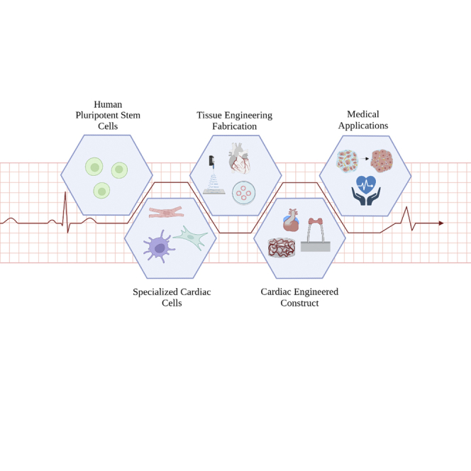
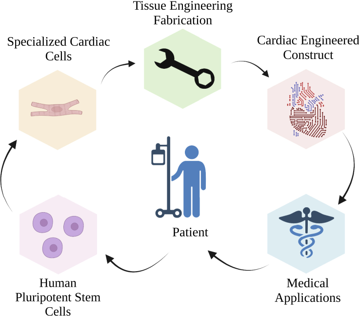
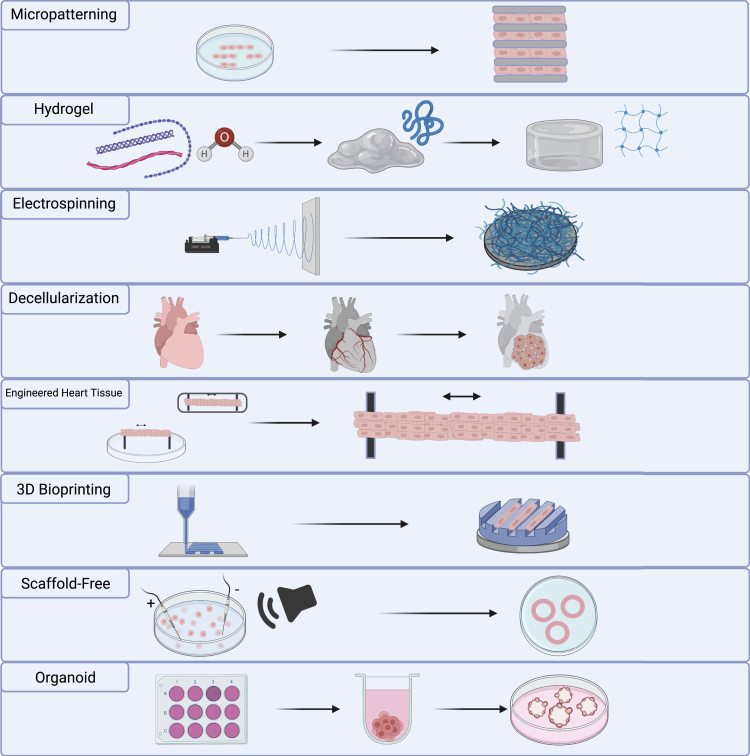
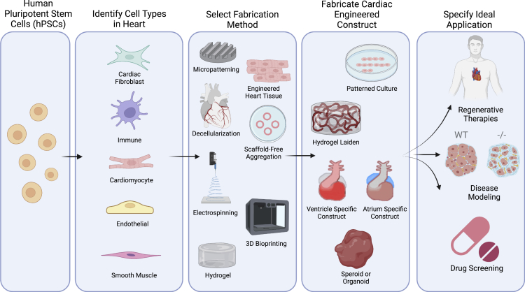
3D cardiac engineered constructs have yielded not only the next generation of cardiac regenerative medicine but also have allowed for more accurate modeling of both healthy and diseased cardiac tissues. This is critical as current cardiac treatments are rudimentary and often default to eventual heart transplants. This review serves to highlight the various cell types found in cardiac tissues and how they correspond with current advanced fabrication methods for creating cardiac engineered constructs capable of shedding light on various pathologies and providing the therapeutic potential for damaged myocardium. In addition, insight is given toward the future direction of the field with an emphasis on the creation of specialized and personalized constructs that model the region-specific microtopography and function of native cardiac tissues.
Keywords: 3d reconstruction of protein; Biomaterials; Materials science.
© 2022 The Author(s).
Conflict of interest statement
The authors declare no competing interests.
Figures
Similar articles
-
Nano-biomaterials and advanced fabrication techniques for engineering skeletal muscle tissue constructs in regenerative medicine.Nano Converg. 2023 Oct 21;10(1):48. doi: 10.1186/s40580-023-00398-y. Nano Converg. 2023. PMID: 37864632 Free PMC article. Review.
-
Engineering Three-Dimensional Vascularized Cardiac Tissues.Tissue Eng Part B Rev. 2022 Apr;28(2):336-350. doi: 10.1089/ten.TEB.2020.0343. Epub 2021 Mar 16. Tissue Eng Part B Rev. 2022. PMID: 33559514 Free PMC article. Review.
-
Vascularization of three-dimensional engineered tissues for regenerative medicine applications.Acta Biomater. 2016 Sep 1;41:17-26. doi: 10.1016/j.actbio.2016.06.001. Epub 2016 Jun 2. Acta Biomater. 2016. PMID: 27262741 Free PMC article. Review.
-
Engineering and Assessing Cardiac Tissue Complexity.Int J Mol Sci. 2021 Feb 2;22(3):1479. doi: 10.3390/ijms22031479. Int J Mol Sci. 2021. PMID: 33540699 Free PMC article. Review.
-
Engineered biomaterials to guide spheroid formation, function, and fabrication into 3D tissue constructs.Acta Biomater. 2023 Jul 15;165:4-18. doi: 10.1016/j.actbio.2022.09.052. Epub 2022 Sep 24. Acta Biomater. 2023. PMID: 36167240 Free PMC article. Review.
Cited by
-
Recent Advances in Hydrogel-Based 3D Bioprinting and Its Potential Application in the Treatment of Congenital Heart Disease.Biomolecules. 2024 Jul 18;14(7):861. doi: 10.3390/biom14070861. Biomolecules. 2024. PMID: 39062575 Free PMC article. Review.
-
The Current State of Realistic Heart Models for Disease Modelling and Cardiotoxicity.Int J Mol Sci. 2024 Aug 24;25(17):9186. doi: 10.3390/ijms25179186. Int J Mol Sci. 2024. PMID: 39273136 Free PMC article. Review.
-
Advances in the Generation of Constructed Cardiac Tissue Derived from Induced Pluripotent Stem Cells for Disease Modeling and Therapeutic Discovery.Cells. 2024 Jan 29;13(3):250. doi: 10.3390/cells13030250. Cells. 2024. PMID: 38334642 Free PMC article. Review.
-
Tissue-Engineered Constructions in Biophysics, Neurology and Other Fields and Branches of Medicine.Biophysics (Oxf). 2022;67(5):816-834. doi: 10.1134/S0006350922050141. Epub 2022 Dec 19. Biophysics (Oxf). 2022. PMID: 36567971 Free PMC article.
-
Suspended Tissue Open Microfluidic Patterning (STOMP).bioRxiv [Preprint]. 2025 Mar 29:2024.10.04.616662. doi: 10.1101/2024.10.04.616662. bioRxiv. 2025. Update in: Adv Sci (Weinh). 2025 Jul;12(25):e2501148. doi: 10.1002/advs.202501148. PMID: 39416011 Free PMC article. Updated. Preprint.
References
-
- Abilez O.J., Tzatzalos E., Yang H., Zhao M.T., Jung G., Zollner A.M., Tiburcy M., Riegler J., Matsa E., Shukla P., et al. Passive stretch induces structural and functional maturation of engineered heart muscle as predicted by computational modeling. Stem Cells. 2018;36:265–277. doi: 10.1002/stem.2732. - DOI - PMC - PubMed
-
- Al-Hejailan R.S., Bakheet R.H., Al-Saud M.M., Al-Jufan M.B., Al-Hindas H.M., Al-Qattan S.M., Al-Muhanna M.K., Parhar R.S., Conca W., Hansmann J., et al. Toward allogenizing a xenograft: xenogeneic cardiac scaffolds recellularized with human-induced pluripotent stem cells do not activate human naïve neutrophils. J. Biomed. Mater. Res. B: Appl. Biomater. 2021;110:691–701. doi: 10.1002/JBM.B.34948. - DOI - PubMed
-
- Allyson Walker C., Francis Spinale B.G. Basic science review the structure and function of the cardiac myocyte: a review of fundamental concepts. J. Thorac. Cardiovasc. Surg. 1999;465:747–763. - PubMed
Publication types
LinkOut - more resources
Full Text Sources
