

Case Reports
doi: 10.1016/j.case.2022.01.010.
eCollection 2022 May.
Membranous Ventricular Septal Aneurysm Leading to Embolic Stroke
Affiliations
- PMID: 35602978
- PMCID: PMC9120855
- DOI: 10.1016/j.case.2022.01.010
Item in Clipboard
Case Reports
Membranous Ventricular Septal Aneurysm Leading to Embolic Stroke
CASE (Phila).
.
No abstract available
Keywords: Embolism; Ischemic stroke; Membranous ventricular septal aneurysm; Ultrasound enhancing agent.
Figures
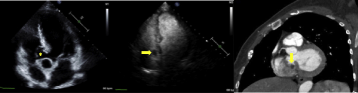
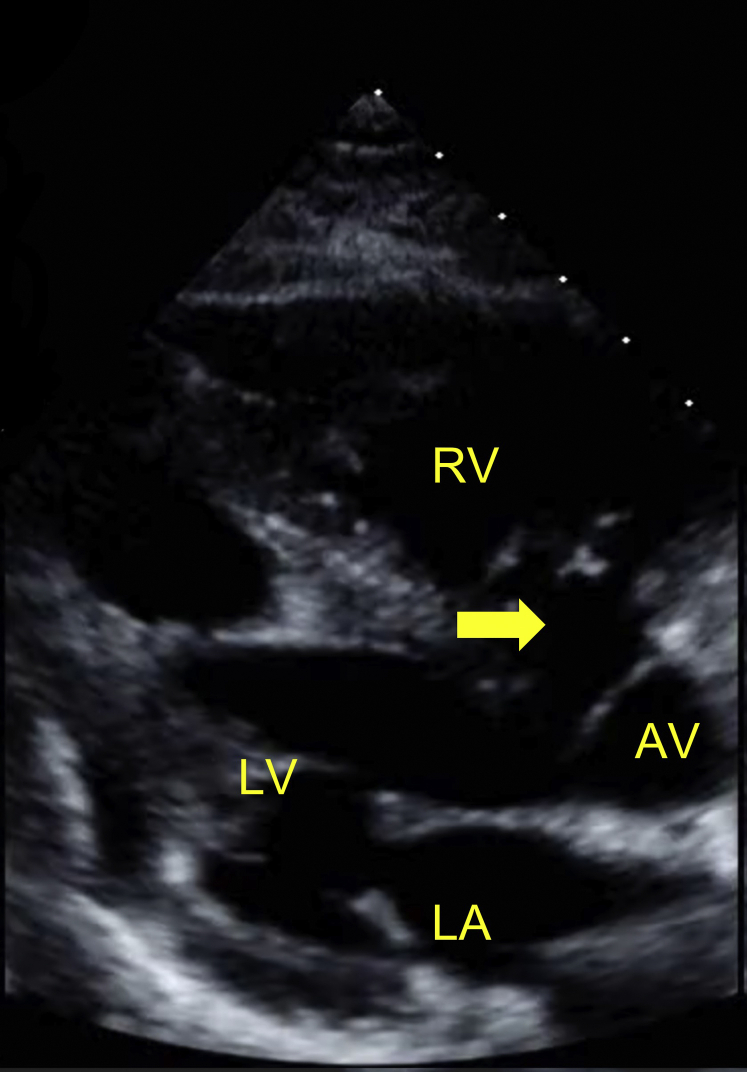
Two-dimensional TTE, modified parasternal long-axis orientation in late-diastolic phase demonstrates the large MVSA (arrow). AV, Aortic valve; LA, left atrium; LV, left ventricle; RV, right ventricle.
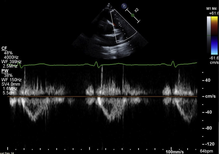
Two-dimensional basal parasternal short-axis view, pulsed-wave Doppler of the RVOT.
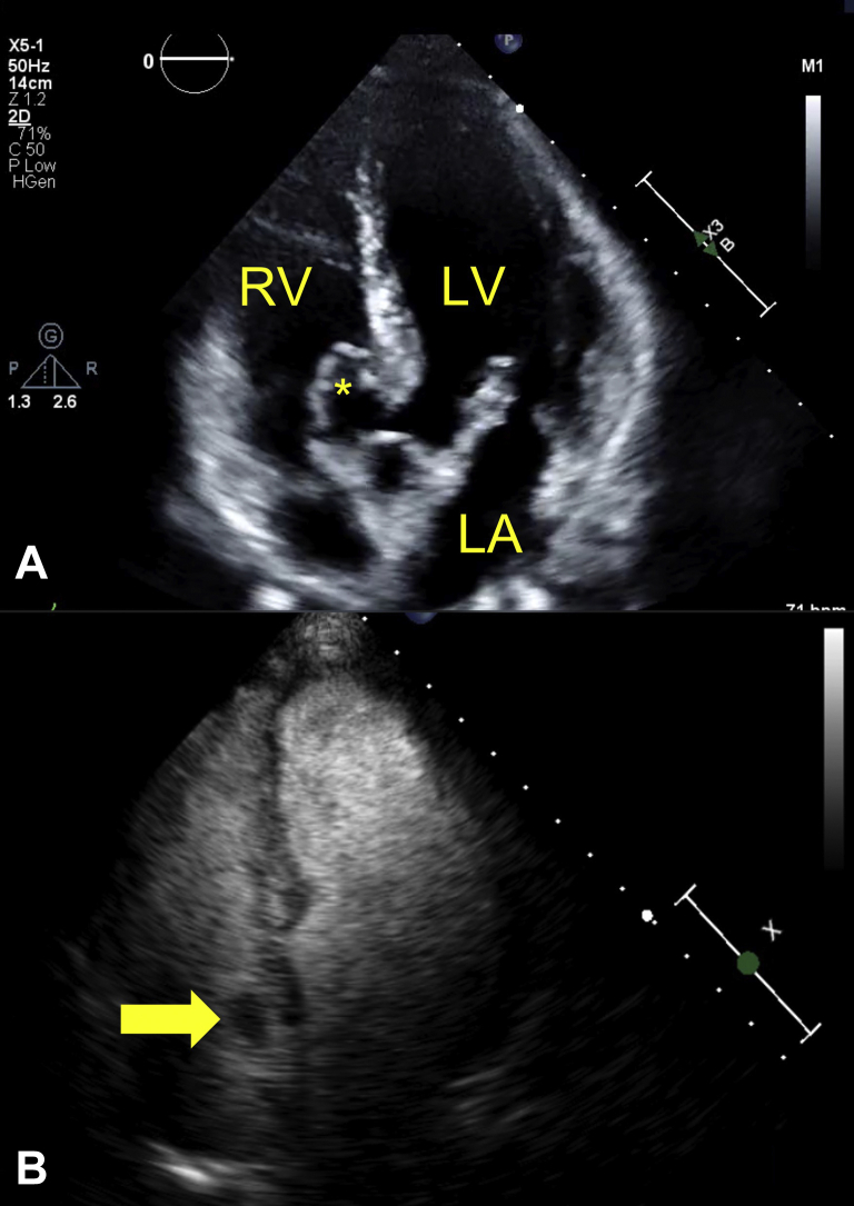
Two-dimensional TTE, apical five-chamber view, diastolic phase without (A) and with (B) an UEA demonstrates the large MVSA (∗) bulging into the RV. A filling defect is seen inside the MVSA (arrow) consistent with a suspected thrombus (B). LA, Left atrium; LV, left ventricle; RV, right ventricle. The arrow indicates the thrombus.

Contrast-enhanced CCT scan, multiplanar reconstruction, coronal display (A), and maximal intensity projection with increased slice thickness, oblique sagittal short-axis display (B). The filling defect (thrombus) is well visualized (arrows) and can be measured at 0.8 × 1.2 cm. Ao, Ascending aorta; AV, aortic valve; LV, left ventricle; PV, pulmonic valve. The arrow indicates the thrombus.
References
-
- Misra P., Hildner F.J., Cohen L.S., Narula O.S., Samet P. Aneurysm of the membranous ventricular septum—a mechanism for spontaneous closure of the VSD. N Engl J Med. 1970;283:58–61. - PubMed
-
- Mangla A., Kalra D.K. Membranous ventricular septum aneurysm, differentiated from sinus of valsalva aneurysm using cardiac CT. J Cardiovasc Comput Tomogr. 2018;12:92–94. - PubMed
-
- Choi M., Jung J.I., Lee B.Y., Kim H.R. Ventricular septal aneurysms in adults: findings of cardiac CT images and correlation with clinical features. Acta Radiol. 2011;52:619–623. - PubMed
-
- Salazar J., Gutierrez A., Cay E., Ballester C., Salazar J.J., Placer L. Cerebral embolism and thrombus in a membranous interventricular septal aneurysm. Ann Thorac Surg. 2003;76:286–287. - PubMed
-
- Lin J.M., Hwang J.J., Chiu I.S. Cerebral embolism from the thrombus in the atrioventricular septal aneurysm. Cardiology. 1995;86:441–443. - PubMed
Publication types
LinkOut - more resources
Full Text Sources
