

Whole slide imaging vs eyeballing: The future in quantification of tubular atrophy in routine clinical practice
- PMID: 35603119
- PMCID: PMC9121712
- DOI: 10.4103/ijn.IJN_333_20
Whole slide imaging vs eyeballing: The future in quantification of tubular atrophy in routine clinical practice
Abstract
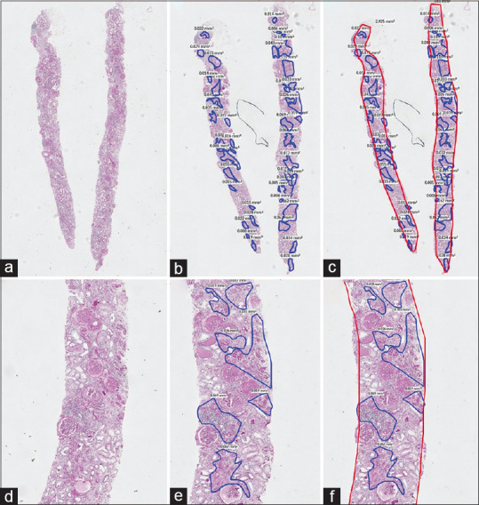
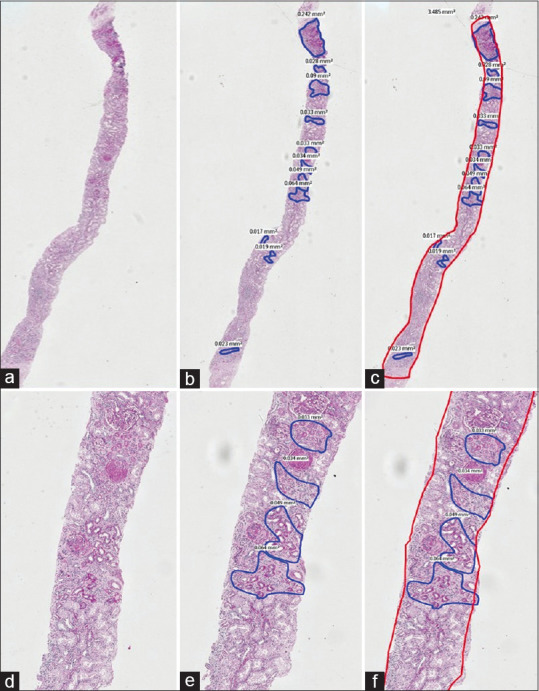
Introduction: Histologic assessment of interstitial fibrosis and tubular atrophy is an accepted method of assessing chronic damage to the kidney and correlates with renal function in native and allograft renal biopsies. The challenge, however, is to quantify the interstitial fibrosis and tubular atrophy with accuracy and to minimize the inter-observer variability. Though "eyeballing" on light microscopy is the most commonly practised method used for the quantification of tubular atrophy, it may not be very accurate. To complement this method, Whole Slide Imaging (WSI) techniques that have more accurate results and have a higher reproducibility can be used. There is not much data on the correlation of the results obtained by the 'eyeballing' technique with those by digital WSI.
Methods: Tubular atrophy in 151 consecutive adequate native kidney biopsies were graded 0 to III by 'conventional' eyeballing by a single experienced renal pathologist. These results were compared with the grades obtained on the same cases by WSI and digital marking of the atrophy.
Results: The concordance of the two groups in the entire cohort was only 66.2% with over grading in 30.4% and under grading in 3.3%. Whilst accuracy of grading was over 74% in all grades, the sensitivity in grades I and II were low at 52% and 47.3% respectively as was the positive predictive value at 32.5 and 44% respectively.
Conclusion: Assessment of tubular atrophy on digital images will be the way forward for accurate quantification.
Keywords: Eyeballing; tubular atrophy; whole slide imaging.
Copyright: © 2022 Indian Journal of Nephrology.
Conflict of interest statement
There are no conflicts of interest.
Figures
References
-
- Sethi S, D’Agati V, Nast CC, Fogo AB, De Vriese AS, Markowitz GS, et al. A proposal for standardized grading of chronic changes in native kidney biopsy specimens. Kidney Int. 2017;91:787–9. - PubMed
-
- Bellur SS, Roberts ISD, Troyanov S, Royal V, Coppo R, Cook HT, et al. Reproducibility of the Oxford classification of immunoglobulin A nephropathy, impact of biopsy scoring on treatment allocation and clinical relevance of disagreements: Evidence from the VALidation of IGA study cohort. Nephrol Dial Transplant. 2019;34:1681–90. - PubMed
-
- Wernick RM, Smith DL, Houghton DC, Phillips DS, Booth JL, Runckel DN, et al. Reliability of histologic scoring for lupus nephritis: A community-based evaluation. Ann Intern Med. 1993;119:805–11. - PubMed
-
- Oni L, Beresford MW, Witte D, Chatzitolios A, Sebire N, Abulaban K, et al. Inter-observer variability of the histological classification of lupus glomerulonephritis in children. Lupus. 2017;26:1205–11. - PubMed
-
- Grootscholten C, Bajema IM, Florquin S, Steenbergen EJ, Peutz-Kootstra CJ, Goldschmeding R, et al. Interobserver agreement of scoring of histopathological characteristics and classification of lupus nephritis. Nephrol Dial Transplant. 2008;23:223–30. - PubMed
LinkOut - more resources
Full Text Sources