Review
. 2022 May 6:13:747799.
doi: 10.3389/fimmu.2022.747799. eCollection 2022.

Macrophage: A Cell With Many Faces and Functions in Tuberculosis

Affiliations
Review

Macrophage: A Cell With Many Faces and Functions in Tuberculosis

Faraz Ahmad et al. Front Immunol. .

Abstract

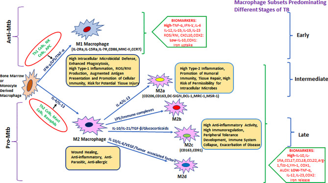
Mycobacterium tuberculosis (Mtb) is the causative agent of human tuberculosis (TB) which primarily infects the macrophages. Nearly a quarter of the world's population is infected latently by Mtb. Only around 5%-10% of those infected develop active TB disease, particularly during suppressed host immune conditions or comorbidity such as HIV, hinting toward the heterogeneity of Mtb infection. The aerosolized Mtb first reaches the lungs, and the resident alveolar macrophages (AMs) are among the first cells to encounter the Mtb infection. Evidence suggests that early clearance of Mtb infection is associated with robust innate immune responses in resident macrophages. In addition to lung-resident macrophage subsets, the recruited monocytes and monocyte-derived macrophages (MDMs) have been suggested to have a protective role during Mtb infection. Mtb, by virtue of its unique cell surface lipids and secreted protein effectors, can evade killing by the innate immune cells and preferentially establish a niche within the AMs. Continuous efforts to delineate the determinants of host defense mechanisms have brought to the center stage the crucial role of macrophage phenotypical variations for functional adaptations in TB. The morphological and functional heterogeneity and plasticity of the macrophages aid in confining the dissemination of Mtb. However, during a suppressed or hyperactivated immune state, the Mtb virulence factors can affect macrophage homeostasis which may skew to favor pathogen growth, causing active TB. This mini-review is aimed at summarizing the interplay of Mtb pathomechanisms in the macrophages and the implications of macrophage heterogeneity and plasticity during Mtb infection.

Keywords: Mycobacterium tuberculosis; innate immunity; macrophage heterogeneity; metabolic reprogramming; phenotype switching; trained immunity.


Conflict of interest statement

The authors declare that the research was conducted in the absence of any commercial or financial relationships that could be construed as a potential conflict of interest.

Figures

Figure 1
Figure 1
Heterogeneity of bone-marrow- or monocyte-derived macrophages and their physiological roles with reference to TB. The fate and function of recruited macrophages are generally shaped with influence from the local environment, stimulatory signals, and type of infection. Effector cells, including Th1/NK cells and APCs, displaying antigenic peptides from intracellular pathogens (or due to stimulation with LPS/IFN-γ), give rise to the M1 type of pro-inflammatory macrophages that either clear or restrain (through granulomatous response) intracellular infections including Mycobacterium tuberculosis (Mtb). M1 macrophages are generally characterized by a high level of pro-inflammatory mediators such as TNF-α, IFN-γ, IL-6, IL-12, IL-1β, IL-15, IL-23, CXCL-10, COX-2, and ROS/RNI and a low level of immune-regulatory molecules including IL-10, IL-4, TGF-β, and COX-1. In parallel, during invasion by an extracellular pathogen, Th2 cells/mast cells/basophils (or stimulation with IL-4, IL-10, IL-13, and immune complexes) act to differentiate macrophages toward the M2 state that generally ensure clearance of extracellular parasites. M2 macrophages are generally characterized by the production of a high level of IL-10, IL-1Ra, CCL17, CCL18, CCL22, Arg-1, fizz-1, Ym-1, COX-1, ALOX, etc. and a low level of TNF-α, IL-12, IL-23, and COX2 among others and further divided into various subtypes such as M2a, M2b, M2c, and the most recently described M2d. They are known for their role in promoting intracellular infections, for example, AMs in Mtb infection.
Figure 2
Figure 2
An immunometabolic circuit that dictates macrophage fate and function in tuberculosis. M1 macrophages that restrict Mtb proliferation are generally glycolytically active and utilize more glucose to meet increased energy demand as a result of enhanced proliferation. M1 macrophages skip the tricarboxylic acid (TCA) cycle and prefer energetically favored lactate production from pyruvate even in the absence of oxygen (Warburg effect) to which support rapid cellular turnover and generation of anti-mycobacterial oxidative burst. Instead of the TCA cycle, they utilize the pentose phosphate pathway and citric acid cycle for extra-mitochondrial utilization of available fatty acids to meet increased energy demands to support activation of pro-inflammatory genes including NF-κβ, IRF-3/5, STAT-1, and HIF-1α and their downstream effectors such as TNF-α, IL-12, IFN-β/γ, CCL-5, and CXCL-4/9/10/11 to restrict Mtb growth. In stark contrast, both AMs and M2 macrophages that support Mtb persistence/proliferation elect cost-inefficient mitochondrial oxidative phosphorylation (OXPHOS) and fatty acid oxidation (FAO) pathways to meet cellular demands. Mtb exploit these pathways to hijack host cells and utilizes the host’s own lipids to thrive in hibernation for longer periods. In M2 macrophages, it induces the cellular expression of anti-inflammatory mediators including STAT-3/6, C-Maf, and HIF-2α that stimulate the production of downstream anti-inflammatory effectors including IL-10, IL-1Rα, IL-4Rα, Arg-1/2, and PPAR-γ among others and thus make a permissive environment for Mtb growth as well persistence.

References

    1. Brites D, Gagneux S. Co-Evolution of Mycobacterium Tuberculosis and Homo Sapiens. Immunol Rev (2015) 264:6–24. doi: 10.1111/imr.12264 - DOI - PMC - PubMed
    1. Comas I, Coscolla M, Luo T, Borrell S, Holt KE, Kato-Maeda M, et al. . Out-Of-Africa Migration and Neolithic Coexpansion of Mycobacterium Tuberculosis With Modern Humans. Nat Genet (2013) 45:1176–82. doi: 10.1038/ng.2744 - DOI - PMC - PubMed
    1. Bos KI, Harkins KM, Herbig A, Coscolla M, Weber N, Comas I, et al. . Pre-Columbian Mycobacterial Genomes Reveal Seals as a Source of New World Human Tuberculosis Provided Samples for Analysis HHS Public Access. Nature (2014) 514:494–7. doi: 19.1038/nature13591 - PMC - PubMed
    1. Sabin S, Herbig A, Vågene ÅJ, Ahlström T, Bozovic G, Arcini C, et al. . A Seventeenth-Century Mycobacterium Tuberculosis Genome Supports a Neolithic Emergence of the Mycobacterium Tuberculosis Complex. Genome Biol (2020) 21:201. doi: 10.1186/s13059-020-02112-1 - DOI - PMC - PubMed
    1. Hershkovitz I, Donoghue HD, Minnikin DE, May H, Lee OY, Feldman M, et al. . Tuberculosis Origin: The Neolithic Scenario. Tuberculosis (2015) 95:S122–6. doi: 10.1016/j.tube.2015.02.021 - DOI - PubMed