Case Reports
. 2022 Mar 15;25(1):32-35.
doi: 10.7602/jmis.2022.25.1.32.

Dome-type carcinoma of the rectum mimicking a submucosal tumor: a case report and literature review

Affiliations
Case Reports

Dome-type carcinoma of the rectum mimicking a submucosal tumor: a case report and literature review

Byeong-Joo Noh et al. J Minim Invasive Surg. .

Abstract

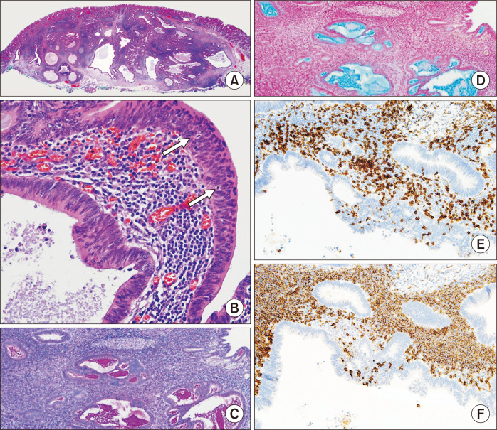
Dome-type carcinoma (DC) has been recognized as a rare variant of adenocarcinoma, which arises in gut-associated lymphoid tissue. It has a specific morphologic feature of a dome-like protrusion associated with lymphoid tissue. We report a case of a DC of the rectum in an asymptomatic 58-year-old male. A 2-cm sized, well-demarcated, round mass masquerading as a submucosal tumor (SMT) was identified in the rectum and was resected by endoscopic submucosal dissection. The tumor was revealed as an adenocarcinoma with submucosal invasion of 3,700 µm, which consisted of dilated cystic glands and the lymphoid stroma with reactive germinal centers. On immunohistochemistry, the tumor cells revealed retained expression for mismatch repair proteins. Laparoscopic surgical resection was subsequently performed. DC is considered a distinctive subtype of colorectal adenocarcinoma with characteristic morphology and low-grade malignant potential. Careful detection of the overlying mucosal lesion is crucial to differentially diagnose DC from SMT.

Keywords: Adenocarcinoma; Carcinoma; Colorectal neoplasms; Lymphoid tissue; Morphology.


Figures

Fig. 1
Fig. 1
(A) Colonoscopy identifies a mucosa-covered, protruding, round mass with shallow erosion, originating from the rectum. (B) Endoscopic ultrasound shows a well-demarcated, heterogeneous, and hyperechoic submucosal mass, emanating from the mucosal layer of the rectum.
Fig. 2
Fig. 2
(A) The microscopic pathological examination reveals a well-delimited expansile tumor, measuring 1.6 × 1.6 × 0.4 cm, invading the submucosa (H&E stain, ×12.5). (B) The neoplastic glands are composed of pseudostratified cuboidal to columnar and mucin-depleted (absence of goblet cells) epithelial cells with eosinophilic cytoplasm and vesicular nuclei. Focal tumor-infiltrating lymphocytes and intraluminal pink material from the neoplastic glands are also noted (H&E stain, ×200). (C, D) The pink material shows intensive periodic acid–Schiff-diastase-positive (C, ×40) and Alcian blue-positive staining (D, ×40). (E, F) The tumor-infiltrating lymphocytes consist of mixed CD3- (E, ×200) and CD20-positive cells (F, ×200).

References

    1. Neutra MR, Mantis NJ, Kraehenbuhl JP. Collaboration of epithelial cells with organized mucosal lymphoid tissues. Nat Immunol. 2001;2:1004–1009. doi: 10.1038/ni1101-1004. - DOI - PubMed
    1. Rubio CA, Lindh C, Björk J, Törnblom H, Befrits R. Protruding and non-protruding colon carcinomas originating in gut-associated lymphoid tissue. Anticancer Res. 2010;30:3019–3022. - PubMed
    1. Rubio CA, Befrits R, Ericsson J. Carcinoma in gut-associated lymphoid tissue in ulcerative colitis: case report and review of literature. World J Gastrointest Endosc. 2013;5:293–296. doi: 10.4253/wjge.v5.i6.293. - DOI - PMC - PubMed
    1. De Petris G, Lev R, Quirk DM, Ferbend PR, Butmarc JR, Elenitoba-Johnson K. Lymphoepithelioma-like carcinoma of the colon in a patient with hereditary nonpolyposis colorectal cancer. Arch Pathol Lab Med. 1999;123:720–724. doi: 10.5858/1999-123-0720-LLCOTC. - DOI - PubMed
    1. Jass JR, Constable L, Sutherland R, et al. Adenocarcinoma of colon differentiating as dome epithelium of gut-associated lymphoid tissue. Histopathology. 2000;36:116–120. doi: 10.1046/j.1365-2559.2000.00864.x. - DOI - PubMed

Publication types

LinkOut - more resources