. 2022 May;13(5):12409-12420.
doi: 10.1080/21655979.2022.2074767.

Wnt family member 1 (Wnt1) overexpression-induced M2 polarization of microglia alleviates inflammation-sensitized neonatal brain injuries

Affiliations

Wnt family member 1 (Wnt1) overexpression-induced M2 polarization of microglia alleviates inflammation-sensitized neonatal brain injuries

Jinzhi Gao et al. Bioengineered. 2022 May.

Abstract

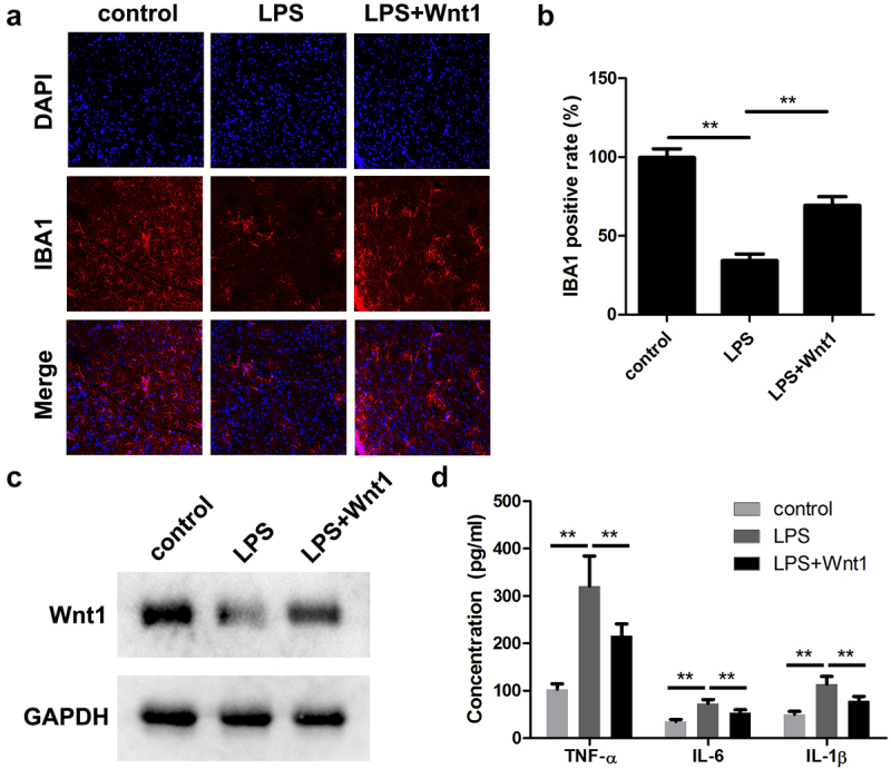
Intrauterine infection induces inflammation-mediated microglial activation and brain injury. This study aimed to explore the regulatory mechanism of Wnt family member 1 (Wnt1) in intrauterine infection-mediated microglial polarization. The cell counting kit-8 (CCK-8) assay was used to determine the viability of microglia, and cytokine expression levels were determined using enzyme linked immunosorbent assay (ELISA) kits and real-time quantitative PCR (RT-qPCR). The number of CD206+ and CD16/32+ cells was determined by flow cytometry. Wnt1 expression was analyzed using western blotting and immunofluorescence. Moreover, an in vivo assay was performed to verify the role of WNT1 in inflammation-sensitized brain injury in newborn mice. Lipopolysaccharide (LPS) exposure resulted in a decrease in microglial cell viability while increasing the expression levels of inflammatory cytokines (TNF-α, IL-6, and IL-1β), simultaneously promoting M1-type microglial conversion. However, these effects were rescued by overexpression of Wnt1, which was expressed less in microglia exposed to LPS in vitro and in vivo. Here, we found that Wnt1 activated the LKB1-AMPK pathway, and the inhibition of LKB1 attenuated the rescue effects of Wnt1. In addition, LPS exposure reduced the autophagy of microglia, and Wnt1 overexpression enhanced the autophagy, but this effect was reversed by treatment with an LKB1 inhibitor. Wnt1 activated LKB1 to suppress inflammation-mediated activation of microglia, promote M2-type microglia conversion via the AMPK pathway, and alleviate inflammation-sensitized neonatal brain injuries. This provides a potential avenue for the treatment of neonatal brain injuries.

Keywords: AMPK; LKB1; Microglia polarization; autophagy; wnt1.


Conflict of interest statement

No potential conflict of interest was reported by the author(s).

Figures

None
Graphical abstract
Figure 1.
Figure 1.
Exposure to LPS promotes M2-to-M1 polarization of microglia. (a) Cell viability of BV2 cells; (b) Levels of TNF-α, IL-6, and IL-1β in BV2 cells; (c) mRNA expression of TNF-α, IL-6, and IL-1β in BV2 cells; (d) Surface expression of CD206 and CD16/32; (e) The percentage of CD16/32 cells; (f) The percentage of CD206 cells. *P < 0.05, **P < 0.01, *** P < 0.001 versus control.
Figure 2.
Figure 2.
Wnt1 protein expression decreases following LPS exposure. (a) mRNA expression of Wnt1 in BV2 cells and culture medium; (b) Protein expression of Wnt1 in BV2 cells and culture medium; (c) Quantification of (B); (d) Immunofluorescence analysis for Wnt1. *P < 0.05, **P < 0.01 versus control.
Figure 3.
Figure 3.
Wnt1 overexpression alters the polarization of microglia. (a) Expression of Wnt1 in BV2 cells and culture medium at mRNA level; (b) Expression of Wnt1 in BV2 cells and culture medium at protein level; (c) Viability of BV2 cells; (d) Levels of TNF-α, IL-6, and IL-1β in BV2 cells; (e) mRNA expression of TNF-α, IL-6, and IL-1β in BV2 cells; (f) Surface expression of CD206 and CD16/32; (g) The percentage of CD16/32 cells; (h) The percentage of CD206 cells. ##P < 0.01, ### P < 0.001 versus LPS + vector. **P < 0.01, *** P < 0.001 versus control.
Figure 4.
Figure 4.
Wnt1 alleviates LPS-induced neonatal brain injuries. (a) The expression of IBA1 was detected using an immunofluorescence assay. (b) Quantification of A. (c) The protein expression of WNT1 was determined using a western blot. (d) The release of TNF-α, IL-6, and IL-1β. **P < 0.01, *** P < 0.001.
Figure 5.
Figure 5.
Wnt1 activates the LKB1-AMPK pathway. (a) Protein expression of Wnt1, p-LKB1, LKB1, p-AMPK, AMPK, and PGC1-α. (b) Quantification of p-LKB1/LKB1 and p-AMPK/AMPK ratio. (c) Quantification of each protein normalized to GAPDH. ###P < 0.001, versus LPS + vector. **P < 0.01, *** P < 0.001 versus control.
Figure 6.
Figure 6.
Wnt1 alters the polarization of microglia via the LKB1-AMPK pathway. (a) Protein expression of Wnt1, p-LKB1, LKB1, p-AMPK, AMPK, and PGC1-α and quantification; (b) Viability of BV2 cells; (c) Levels of TNF-α, IL-6, and IL-1β in BV2 cells; (d) mRNA expression of TNF-α, IL-6, and IL-1β in BV2 cells; (e) Surface expression of CD206 and CD16/32; (f) The percentage of CD16/32 cells; (g) The percentage of CD206 cells. &P < 0.05, &&P < 0.01, &&& P < 0.001 versus LPS + Wnt1. ##P < 0.01, ### P < 0.001 versus LPS. *** P < 0.001 versus control.
Figure 7.
Figure 7.
Wnt1 reduces the suppression of autophagy in microglia by LPS. (a) Protein expression of LC3 I, LC3 II, Beclin 1, and P62; (b) Quantification of (A); (c) GFP-tagged LC3 in BV2 cells. # P < 0.05 versus LPS + Wnt1. *** P < 0.001 versus control.

Similar articles

Cited by

References

    1. Fu CH, Zhang BH, Fang CZ, et al. Long non-coding RNA CRNDE deteriorates intrauterine infection-induced neonatal brain injury. Mol Cell Probes. 2020;52:101565. - PubMed
    1. Vermillion MS, Lei J, Shabi Y, et al. Intrauterine Zika virus infection of pregnant immunocompetent mice models transplacental transmission and adverse perinatal outcomes. Nat Commun. 2017;8(1):14575. - PMC - PubMed
    1. Yuan TM, Sun Y, Zhan CY, et al. Intrauterine infection/inflammation and perinatal brain damage: role of glial cells and Toll-like receptor signaling. J Neuroimmunol. 2010;229(1–2):16–25. - PubMed
    1. Hester MS, Tulina N, Brown A, et al. Intrauterine inflammation reduces postnatal neurogenesis in the hippocampal subgranular zone and leads to accumulation of hilar ectopic granule cells. Brain Res. 2018;1685:51–59. - PMC - PubMed
    1. Fan LW, Pang Y.. Dysregulation of neurogenesis by neuroinflammation: key differences in neurodevelopmental and neurological disorders. Neural Regen Res. 2017;12(3):366–371. - PMC - PubMed