Review
. 2022 Oct;35(5):545-562.
doi: 10.1177/19714009221100983. Epub 2022 May 22.

Autoimmune disease of head and neck, imaging, and clinical review

Affiliations
Review

Autoimmune disease of head and neck, imaging, and clinical review

Alireza Paydar et al. Neuroradiol J. 2022 Oct.

Abstract

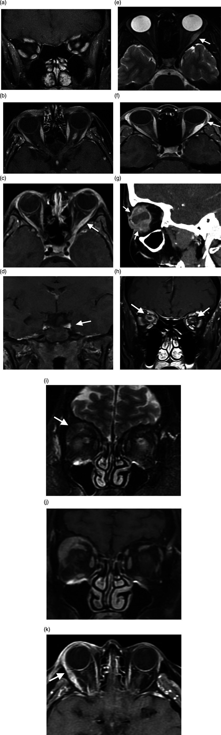
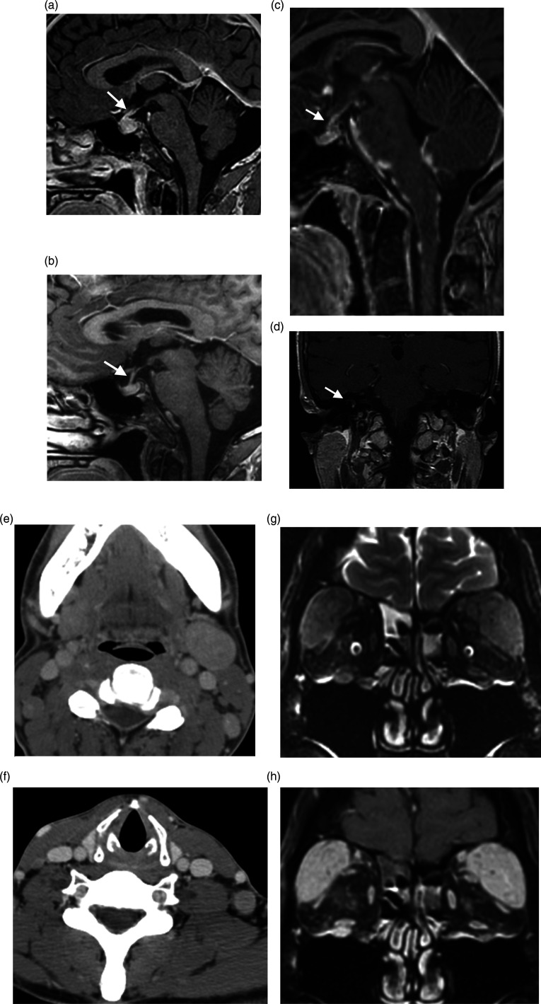
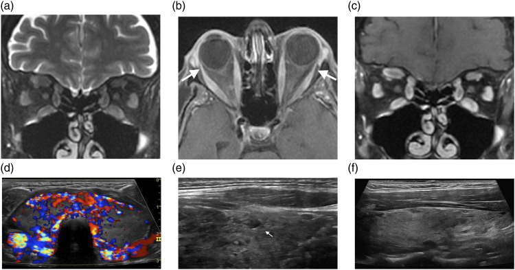
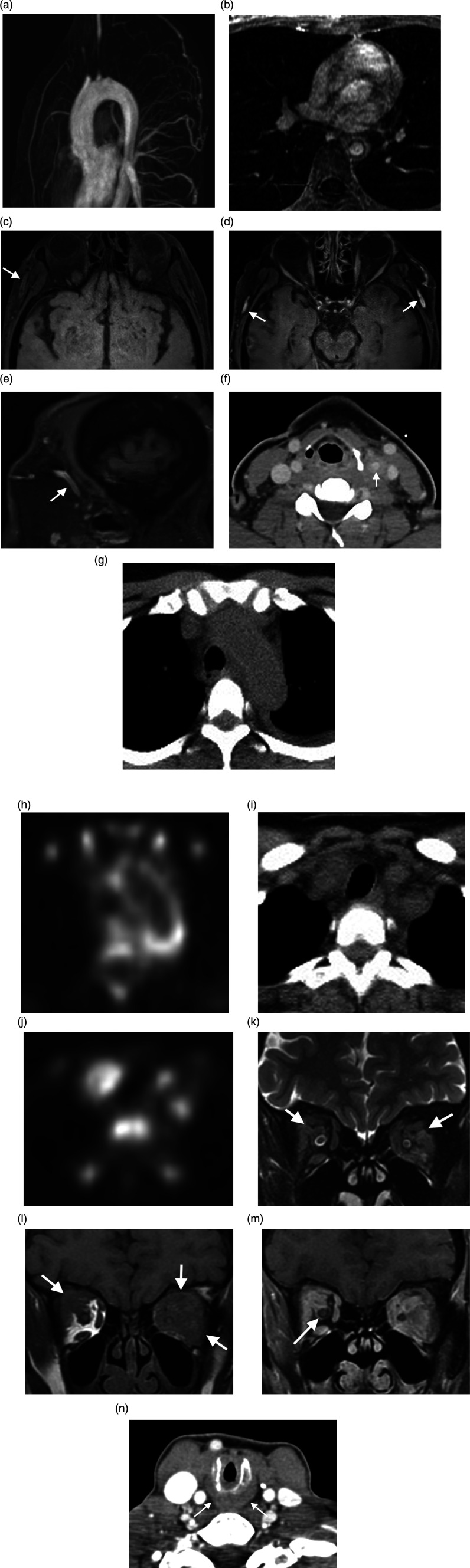
Autoimmune disease of the head and neck (H&N) could be primary or secondary to systemic diseases, medications, or malignancies. Immune-mediated diseases of the H&N are not common in daily practice of radiologists; the diagnosis is frequently delayed because of the non-specific initial presentation and lack of familiarity with some of the specific imaging and clinical features. In this review, we aim to provide a practical diagnostic approach based on the specific radiological findings for each disease. We hope that our review will help radiologists expand their understanding of the spectrum of the discussed disease entities, help them narrow the differential diagnosis, and avoid unnecessary tissue biopsy when appropriate based on the specific clinical scenarios.

Keywords: Autoimmune disease; Head and neck; Head and neck autoimmune disease; Imaging.


Conflict of interest statement

Declaration of conflicting interests: The author(s) declared no potential conflicts of interest with respect to the research, authorship, and/or publication of this article.

Figures

Figure 1.
Figure 1.
The Orbit. (A, B). Neuromyelitis optica. Mild enhancement and edema in the left retrobulbar optic nerve on coronal and axial contrast-enhanced T1WI (A, B). The Orbit. (C, D) Tolosa-Hunt Syndrome. Axial (C) and coronal (D) contrast-enhanced T1WI demonstrates left lateral rectus muscle enlargement and enhancement (arrow, C) which extends to the orbital apex and cavernous sinus (arrow, D). The Orbit. (E, F) Orbital inflammatory pseudotumor. Enlarged and edematous left lateral rectus muscle on axial T2WI (arrow, E) demonstrates homogenous enhancement which spares the insertion on axial contrast-enhanced T1WI (arrow, F). The Orbit. (G) Uveitis. Sagittal contrast-enhanced CT soft tissue window image of the face demonstrates globe irregularity, marked irregular thickening/enhancement of the uvea and sclera (arrows), inhomogeneous vitreous density, and globe rupture with a small air focus (not shown). The Orbit. (H) Optic perineuritis. Coronal contrast-enhanced T1WI shows right greater than left retro-orbital optic nerve sheath enhancement (arrows). The Orbit. (I–K) Lacrimal gland inflammation. Coronal T2WI demonstrates right lacrimal gland enlargement (arrow, I), which enhances on coronal (J) and axial (K) contrast-enhanced fat sat T1WI; adjacent periorbital stranding and enhancement of the right lateral rectus muscle (arrow, K) related to local inflammation.
Figure 2.
Figure 2.
Miscellaneous Head and Neck Diseases. (A, B) Immune checkpoint inhibitor hypophysitis. Sagittal contrast enhanced T1WI demonstrates pituitary gland enlargement and infundibular thickening (arrow, A) in a patient on immunologic therapy for melanoma (ipilimumab and nivolumab), which resolved following medication cessation (arrow, B). Miscellaneous Head and Neck Diseases. (C) Neurosarcoidosis. Sagittal contrast-enhanced fat sat T1WI shows nodular thickening and enhancement of the brainstem leptomeninges and infundibulum (arrow). Miscellaneous Head and Neck Diseases. (D) Cogan Syndrome. Right inner ear saccule enhancement on coronal contrast enhanced T1WI (arrow). Miscellaneous Head and Neck Diseases. (E–H) Castleman Disease. Axial contrast-enhanced neck CT shows cervical lymphadenopathy at the level II (E) and V (F) stations. In a separate patient, lacrimal gland enlargement and enhancement on coronal T2WI (G) and coronal contrast-enhanced T1WI (H), causing exophthalmos. Also note infraorbital nerve enhancement.
Figure 3.
Figure 3.
The Thyroid. (A–D) Graves ophthalmopathy. Coronal T2WI (A) shows diffuse enlargement and edema of the extraocular muscles, which enhance on axial (B) and coronal (C) contrast-enhanced T1WI; mild proptosis and relative sparing of the orbital muscular insertions (arrows, B), consistent with Graves' ophthalmopathy. On transverse color Doppler sonography (D), the gland is diffusely enlarged and hyperemic. The Thyroid. (E) Hashimoto’s thyroiditis. Longitudinal grayscale sonographic image shows glandular enlargement and heterogenous echogenicity; note a small hypoechoic nodule surrounded by echogenic fibrous septa (arrow, E). The Thyroid. (F) Postpartum thyroiditis. Longitudinal grayscale sonographic image shows diffuse hypoechoic glandular parenchyma.
Figure 4.
Figure 4.
Vascular and Inflammatory Diseases. (A, B) Takayasu’s arteritis. MR angiography (A) and axial contrast-enhanced T1WI (B) show descending thoracic aorta circumferential wall thickening and enhancement, consistent with active inflammation. Severe narrowing with smooth tapering in the aortic arch branch vessel ostia. Vascular and Inflammatory Diseases. (C–E) Giant cell arteritis. Vessel wall axial pre-contrast fat sat T1WI demonstrates superficial temporal artery long segment symmetric mural thickening (arrow, C), with vessel wall enhancement on axial and sagittal contrast-enhanced sequences (arrow, D, E). Vascular and Inflammatory Diseases. (F–J) Paraneoplastic carotid arteritis. Axial contrast-enhanced neck CT shows left common carotid artery circumferential wall thickening and perivascular fat stranding (arrow, F). Axial PET/CT shows abnormal FDG avidity in the aortic arch (G, H) and branch vessels (I, J) related to paraneoplastic arteritis in the setting of myelodysplastic syndrome / myeloproliferative disorder. Vascular and Inflammatory Diseases. (K–N) Granulomatosis with polyangiitis (Wegener’s). Coronal T2WI (K) and coronal T1WI (L) show intra-orbital hypointense infiltrating mass replacing the intraconal fat (arrows), which avidly enhances on coronal contrast-enhanced fat sat T1WI (M). While inflammation is contiguous with the extraocular fat and extraocular muscles, the optic nerve sheath complex is preserved (arrow). Axial contrast-enhanced neck CT shows subglottic mucosal edema and wall thickening (arrow, N).

Similar articles

Cited by

  • Autoimmune diseases of the spine and spinal cord.
    Biddle G, Beck RT, Raslan O, Ebinu J, Jenner Z, Hamer J, Hacein-Bey L, Apperson M, Ivanovic V. Biddle G, et al. Neuroradiol J. 2024 Jun;37(3):285-303. doi: 10.1177/19714009231187340. Epub 2023 Jul 3. Neuroradiol J. 2024. PMID: 37394950 Free PMC article. Review.

References

    1. Akaishi T, Nakashima I, Sato DK, et al. Neuromyelitis Optica Spectrum Disorders. Neuroimaging Clin North America. 2017;27(2):251–265. Available from: https://linkinghub.elsevier.com/retrieve/pii/S1052514916301344 - PubMed
    1. Marignier R, Hacohen Y, Cobo-Calvo A, et al. Myelin-oligodendrocyte glycoprotein antibody-associated disease. Lancet Neurol 2021; 20(9): 762–772. - PubMed
    1. Weinshenker BG, Wingerchuk DM. Neuromyelitis Spectrum Disorders. Mayo Clinic Proc 2017; 92(4): 663–679. - PubMed
    1. Ramanathan S, Prelog K, Barnes EH, et al. Radiological differentiation of optic neuritis with myelin oligodendrocyte glycoprotein antibodies, aquaporin-4 antibodies, and multiple sclerosis. Mult Scler J 2016; 22(4): 470–482. - PubMed
    1. Dutra BG, da Rocha AJ, Nunes RH, et al. Neuromyelitis optica spectrum disorders: Spectrum of MR imaging findings and their differential diagnosis. Radiographics 2018; 38(1). - PubMed

MeSH terms