Review
. 2022 May 23;79(6):313.
doi: 10.1007/s00018-022-04332-z.

Mechanisms involved in controlling RNA virus-induced intestinal inflammation

Affiliations
Review

Mechanisms involved in controlling RNA virus-induced intestinal inflammation

Evan Zhang et al. Cell Mol Life Sci. .

Abstract

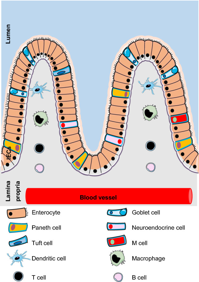
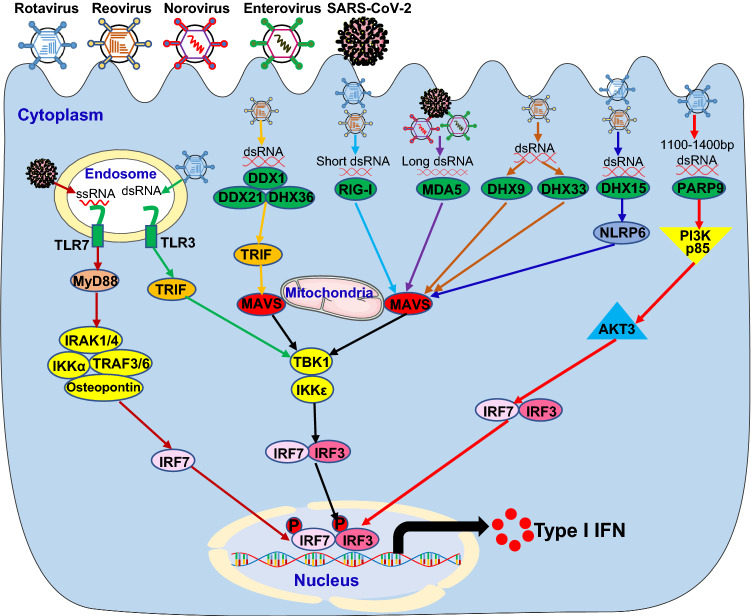
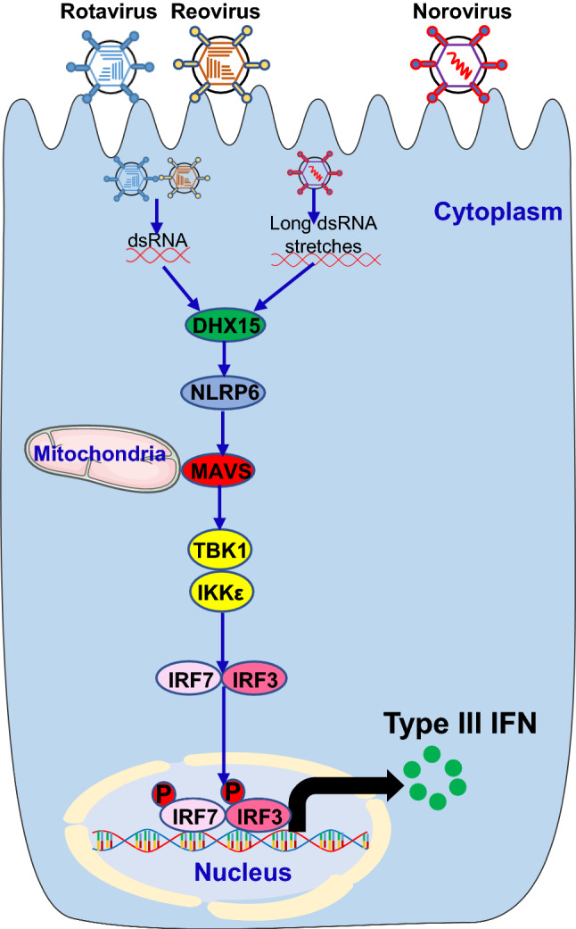
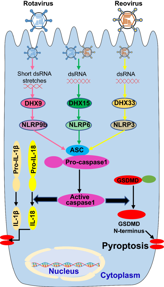
Gastroenteritis is inflammation of the lining of stomach and intestines and causes significant morbidity and mortality worldwide. Many viruses, especially RNA viruses are the most common cause of enteritis. Innate immunity is the first line of host defense against enteric RNA viruses and virus-induced intestinal inflammation. The first layer of defense against enteric RNA viruses in the intestinal tract is intestinal epithelial cells (IECs), dendritic cells and macrophages under the intestinal epithelium. These innate immune cells express pathogen-recognition receptors (PRRs) for recognizing enteric RNA viruses through sensing viral pathogen-associated molecular patterns (PAMPs). As a result of this recognition type I interferon (IFN), type III IFN and inflammasome activation occurs, which function cooperatively to clear infection and reduce viral-induced intestinal inflammation. In this review, we summarize recent findings about mechanisms involved in enteric RNA virus-induced intestinal inflammation. We will provide an overview of the enteric RNA viruses, their RNA sensing mechanisms by host PRRs, and signaling pathways triggered by host PRRs, which shape the intestinal immune response to maintain intestinal homeostasis.

Keywords: Caspase; Cytokine; Diarrhea; Enterocyte; PARP9; Pyroptosis; RNA helicase; SARS-CoV-2.


Conflict of interest statement

The authors declare no conflict of interests.

Figures

Fig. 1
Fig. 1
Overview of IECs and immune cells in intestine. The first layer of host defense against enteric RNA viruses in the intestinal tract is intestinal epithelial cells (IECs), which conclude six cell lineages coming from a common stem cell progenitor: enterocytes, goblet cells, Paneth cells, enteroendocrine cells, tuft cells and microfold (M) cells. Underneath the intestinal epithelium, there is the lamina propria containing the immunocompetent cells infiltrated from blood vessel, which include innate immune cells such as dendritic cells (DCs), macrophages and adaptive immune cells such as T cell and B cells
Fig. 2
Fig. 2
Host PRRs sense enteric RNA viruses to induce type I IFN signaling pathway. Invasion by enteric RNA viruses, including rotavirus, reovirus, norovirus, enterovirus and SARS-CoV-2, introduces RNA into the endosome or dsRNA in the cytoplasm. The host pattern recognition receptors (PRRs), including TLR3 and TLR7 in the endosome and DDX1/DDX21/DHX36 complex, RIG-I, MDA5, DHX9, DHX33, DHX15 and PARP9 in the cytoplasm, recognize the RNA molecules and trigger the activation of downstream cascades through their adaptors leading to the induction of type I IFN in innate immune cells of the intestinal tract. MyD88 Myeloid differentiation primary response 88, TRIF TIR-domain-containing adapter-inducing interferon-β, IRAK1/4 Interleukin 1 receptor-associated kinase 1 and 4, IKKα IκB kinase α, TRAF3/6 TNF receptor-associated factor 3 and 6, IRF7 Interferon regulatory factor 7, DDX1 DEAD-Box helicase 1, RIG-I Retinoic acid-inducible gene I, MDA5 Melanoma differentiation-associated protein 5, DHX15 DEAH-Box helicase 15, PARP9 Poly (ADP-ribose) polymerase 9, MAVS Mitochondrial antiviral-signaling protein, TBK1 TANK-binding kinase 1, NLRP6 the NACHT, LRR, and PYD domains-containing protein 6, PI3K p85 Phosphatidylinositol 3-kinase (PI3K) regulatory subunit p85, AKT3 AKT Serine/Threonine kinase 3
Fig. 3
Fig. 3
The RNA helicase RNA sensor DHX15 sense enteric RNA viruses to trigger type III IFN signaling pathway. Infection by enteric RNA viruses, including rotavirus, reovirus and norovirus, releases dsRNA in the cytoplasm. The RNA helicase RNA sensor DHX15 in the cytoplasm recognizes the RNA molecules and trigger the activation of downstream cascades through their adaptors leading to the induction of type III IFN in IECs of the intestinal tract
Fig. 4
Fig. 4
Host PRRs sense enteric RNA viruses to induce inflammasome activation signaling pathway. Entering by enteric RNA viruses, including rotavirus and reovirus, introduces dsRNA in the cytoplasm. The host PRRs in the cytoplasm, including DHX9, DHX15 and DHX33, recognize the RNA molecules and trigger the activation of inflammasome containing ASC (apoptosis-associated speck-like protein containing a caspase recruitment domain), pro-caspase1 and NLRP9b (the NACHT, LRR, and PYD domains-containing protein 9b), NLRP6 or NLRP3, which results in cleavage of pro-caspase1 into active caspase1. The active caspase 1 mediates maturation and secretion of IL-18 and IL-1β cytokines by cleavage of pro-IL-18 and pro-IL-1β. Additionally, active caspase 1 can provoke a lytic form of cell death termed pyroptosis through cleavage of its substrate Gasdermin D (GSDMD) into GSDMD N-terminus that forms pores in the cellular membrane

Similar articles

Cited by

References

    1. Bányai K, et al. Viral gastroenteritis. Lancet. 2018;392(10142):175–186. - PMC - PubMed
    1. Black RE, et al. Global, regional, and national causes of child mortality in 2008: a systematic analysis. Lancet. 2010;375(9730):1969–1987. - PubMed
    1. Kapikian AZ, et al. Visualization by immune electron microscopy of a 27-nm particle associated with acute infectious nonbacterial gastroenteritis. J Virol. 1972;10(5):1075–1081. - PMC - PubMed
    1. Kapusinszky B, et al. Nearly constant shedding of diverse enteric viruses by two healthy infants. J Clin Microbiol. 2012;50(11):3427–3434. - PMC - PubMed
    1. Jones DL, et al. Shedding of SARS-CoV-2 in feces and urine and its potential role in person-to-person transmission and the environment-based spread of COVID-19. Sci Total Environ. 2020;749:141364. - PMC - PubMed

Substances