. 2022 May 23;79(6):312.
doi: 10.1007/s00018-022-04336-9.

In vitro inhibition of cancer angiogenesis and migration by a nanobody that targets the orphan receptor Tie1

Affiliations

In vitro inhibition of cancer angiogenesis and migration by a nanobody that targets the orphan receptor Tie1

May Meltzer et al. Cell Mol Life Sci. .

Abstract

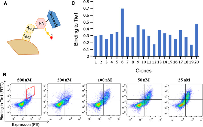
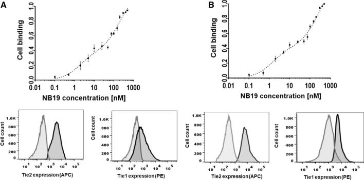
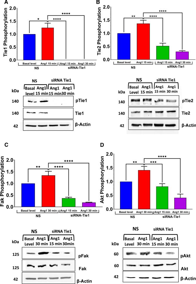
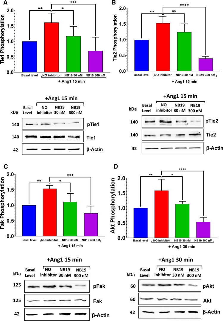
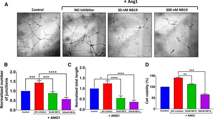
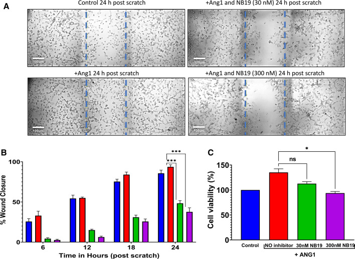
The human signaling molecules Tie1 and Tie2 receptor tyrosine kinases (RTKs) play important pathophysiological roles in many diseases, including different cancers. The activity of Tie1 is mediated mainly through the downstream angiopoietin-1 (Ang1)-dependent activation of Tie2, rendering both Tie 1 and the Tie1/Tie2/Ang1 axis attractive putative targets for therapeutic intervention. However, the development of inhibitors that target Tie1 and an understanding of their effect on Tie2 and on the Tie1/Tie2/Ang1 axis remain unfulfilled tasks, due, largely, to the facts that Tie1 is an orphan receptor and is difficult to produce and use in the quantities required for immune antibody library screens. In a search for a selective inhibitor of this orphan receptor, we sought to exploit the advantages (e.g., small size that allows binding to hidden epitopes) of non-immune nanobodies and to simultaneously overcome their limitations (i.e., low expression and stability). We thus performed expression, stability, and affinity screens of yeast-surface-displayed naïve and predesigned synthetic (non-immune) nanobody libraries against the Tie1 extracellular domain. The screens yielded a nanobody with high expression and good affinity and specificity for Tie1, thereby yielding preferential binding for Tie1 over Tie2. The stability, selectivity, potency, and therapeutic potential of this synthetic nanobody were profiled using in vitro and cell-based assays. The nanobody triggered Tie1-dependent inhibition of RTK (Tie2, Akt, and Fak) phosphorylation and angiogenesis in endothelial cells, as well as suppression of human glioblastoma cell viability and migration. This study opens the way to developing nanobodies as therapeutics for different cancers associated with Tie1 activation.

Keywords: Angiogenesis; Cell migration; Nanobody; Protein engineering; Receptor tyrosine kinase; Tie1.


Conflict of interest statement

The authors declare that they have no competing interests.

Figures

Fig. 1
Fig. 1
Screening of the computationally designed Predes NBlib library for binding to Tie1. a Schematic representation of the Predes NBlib library in the YSD format. b Analysis of nanobody library expression and binding to 100 nM Tie1 after sorting against Tie1 at concentrations of 500, 200, 100, 50, and 25 nM (indicated above each panel). The sorting gate for the first sort is shown in red, and gates of the other sorts were identical to the gate shown in (a). c Binding to Tie1 normalized to expression levels of 20 randomly selected nanobody clones from the fifth sort. The Tie1-binding fluorescence signal for each nanobody clone was divided by its expression signal
Fig. 2
Fig. 2
Binding of NB19 to recombinant and overexpressed Tie1 and Tie2. Representative SPR sensorgrams for binding of Tie1–ECD (a) and Tie2–ECD (b) to immobilized NB19. Different concentrations of Tie1–ECD and Tie2–ECD were allowed to flow over immobilized NB19 for 400 s, and dissociation was monitored for 60 min. c Cell surface expression analysis of Tie2 and Tie1 in transfected vs. untransfected HEK293 cells. HEK293 cells were stained with APC-labeled anti-human Tie2 and PE-labeled anti-human Tie1 antibodies and incubated at 4 °C for 30 min before analysis by flow cytometry. d Affinity titration curves of NB19 binding to HEK293 cells overexpressing Tie1, Tie2, or both Tie1 and Tie2. e Confocal fluorescence microscopy images showing colocalization of NB19 and Tie1 and Tie2 receptors in HEK293 cells overexpressing these receptors. Cell surface expression of Tie2 (green-488) and Tie1 (yellow-PE) are shown; the binding of NB19 was detected by APC anti-his antibody
Fig. 3
Fig. 3
NB19 binding to cells endogenously expressing Tie1 and Tie2. Top panel: Affinity titration curves of NB19 binding to U87-MG cells (a) and TIME cells (b). Two-phase binding curves are shown with R2 = 0.98 and 0.97, respectively. Bottom panel: Cell surface expression of Tie2 and Tie1 in stained (dark gray) U87-MG cells (a) and TIME (b) cells is presented in comparison with unstained cells (light gray). In both cases, cells were stained with APC-labeled anti-human Tie2 antibody and PE-labeled anti-human Tie1 antibody and incubated at 4 °C for 30 min before analysis by flow cytometry
Fig. 4
Fig. 4
Tie1-dependent inhibition of phosphorylation of RTKs in TIME cells. a Determination of Tie1 expression and phosphorylation under different conditions: TIME cells were transfected with siRNA-Tie1 or scrambled siRNA (designated NS) and then treated with control buffer (basal level) or 500 ng/mL Ang1 for 15 or 30 min in a 12-well Multidish plate. Cell lysates were analyzed by western blot using antibodies against phosphorylated Tie1 (pTie1), Tie1, and β-actin. bd Phosphorylation of Tie2, Fak, and Akt, respectively, was determined as in panel (a). In each case, cell lysates were analyzed by western blot using antibodies against the relevant phosphorylated kinase (pTie2, pFak, or pAkt), the kinase itself (Tie2, Fak, or Akt), and β-actin. The intensity of each phosphorylated band measured by ImageJ software was normalized to the expression of total protein of each experiment, and this value was subsequently normalized to the total quantity of β-actin for each sample. Error bars represent SD. One-way ANOVA with Dunnett’s multiple comparison to the Ang1-only treatment was utilized for statistical analysis; *p < 0.1, **p < 0.01, ***p < 0.001, ****p < 0.0001. NS, non-silenced
Fig. 5
Fig. 5
Inhibition of Tie1 and Tie2 phosphorylation by NB19 in TIME cells. a Determination of Tie1 phosphorylation: TIME cells were treated with buffer (control, basal level), 500 ng/mL Ang1, a combination of 500 ng/mL Ang1 and 30 nM/300 nM NB19 for 15 min in a 12-well Multidish plate. Cell lysates were analyzed by western blot using antibodies against phosphorylated Tie1 (pTie1), Tie1, or β-actin. bd Phosphorylation of Tie2, Fak, and Akt, respectively, was determined as in A, with the exception that the incubation time for Akt was 30 min. Cell lysates were analyzed by western blot using antibodies against the relevant phosphorylated kinase (pTie2, pFAK, or pAkt), the kinase itself (Tie2, Fak, or Akt), or β-actin. The intensities of the phosphorylated bands measured by ImageJ were normalized to the expression levels of total protein in each sample, and this value was subsequently normalized to the total quantity of β-actin for each sample. Error bars represent SD. One-way ANOVA with Dunnett’s multiple comparison to the Ang1 treatment was utilized for statistical analysis; *p < 0.1, **p < 0.01, ***p < 0.001, ****p < 0.0001. ns, non-significant
Fig. 6
Fig. 6
Inhibition of tube formation in endothelial cells by NB19. a TIME cells were seeded at 1 × 105 in 8-well plates coated with Matrigel and imaged using confocal microscopy following 18 h of treatment under the conditions indicated in the top panels. Scale bar, 100 µm. b, c Tube structures were analyzed for the number of generated junctions and the total tube length following the different treatments. *p < 0.05; ***p < 0.001; ****p < 0.0001, n = 3. d Effects of NB19 on the growth and survival of TIME cells, as reflected in the number of viable cells assessed by an XTT assay. The results for the different samples were normalized to the untreated control. *p < 0.1, **p < 0.01, ***p < 0.001, ****p < 0.0001. n = 3
Fig. 7
Fig. 7
NB19 suppresses cancer cell migration. A scratch assay was performed on U87-MG cells in the presence of 0, 30, or 300 nM NB19. a Representative photomicrographs of untreated and NB19-treated cells at 24 h post scratch. Broken blue lines indicate the migration front of cells. b Percentage of scratch closure at indicated time points during the assay. The bars are color coded as follows: blue—untreated control cells, red—Ang1-treated cells, green—cells treated with 30 nM NB19 and Ang1, and purple—cells treated with 300 nM NB19 and Ang1. Error bars represent means ± SD of three scratched areas imaged per condition. ***p < 0.001. c Effects of NB19 on the growth and survival of U87-MG cells assessed by an XTT assay. The results for all samples were normalized to the untreated control. *p < 0.05, ns, non-significant. n = 3

Similar articles

Cited by

References

    1. Kontos CD, Cha EH, York JD, Peters KG. The endothelial receptor tyrosine kinase Tie1 activates phosphatidylinositol 3-kinase and Akt to inhibit apoptosis. Mol Cell Biol. 2002;22:1704–1713. doi: 10.1128/mcb.22.6.1704-1713.2002. - DOI - PMC - PubMed
    1. Ward NL, Dumont DJ. The angiopoietins and Tie2/Tek: adding to the complexity of cardiovascular development. Semin Cell Dev Biol. 2002;13:19–27. doi: 10.1006/scdb.2001.0288. - DOI - PubMed
    1. Yancopoulos GD, Davis S, Gale NW, et al. Vascular-specific growth factors and blood vessel formation. Nature. 2000;407:242–248. doi: 10.1038/35025215. - DOI - PubMed
    1. Leppänen V-M, Saharinen P, Alitalo K. Structural basis of Tie2 activation and Tie2/Tie1 heterodimerization. Proc Natl Acad Sci. 2017;114:4376–4381. doi: 10.1073/PNAS.1616166114. - DOI - PMC - PubMed
    1. Savant S, La Porta S, Budnik A, et al. The orphan receptor Tie1 controls angiogenesis and vascular remodeling by differentially regulating Tie2 in tip and stalk cells. Cell Rep. 2015;12:1761–1773. doi: 10.1016/J.CELREP.2015.08.024. - DOI - PMC - PubMed