Functional Alterations Due to COVID-19 Lung Lesions-Lessons From a Multicenter V/Q Scan-Based Registry
- PMID: 35605049
- PMCID: PMC9275799
- DOI: 10.1097/RLU.0000000000004261
Functional Alterations Due to COVID-19 Lung Lesions-Lessons From a Multicenter V/Q Scan-Based Registry
Abstract
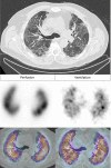
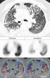
Purpose: In coronavirus disease 2019 (COVID-19) patients, clinical manifestations as well as chest CT lesions are variable. Lung scintigraphy allows to assess and compare the regional distribution of ventilation and perfusion throughout the lungs. Our main objective was to describe ventilation and perfusion injury by type of chest CT lesions of COVID-19 infection using V/Q SPECT/CT imaging.
Patients and methods: We explored a national registry including V/Q SPECT/CT performed during a proven acute SARS-CoV-2 infection. Chest CT findings of COVID-19 disease were classified in 3 elementary lesions: ground-glass opacities, crazy-paving (CP), and consolidation. For each type of chest CT lesions, a semiquantitative evaluation of ventilation and perfusion was visually performed using a 5-point scale score (0 = normal to 4 = absent function).
Results: V/Q SPECT/CT was performed in 145 patients recruited in 9 nuclear medicine departments. Parenchymal lesions were visible in 126 patients (86.9%). Ground-glass opacities were visible in 33 patients (22.8%) and were responsible for minimal perfusion impairment (perfusion score [mean ± SD], 0.9 ± 0.6) and moderate ventilation impairment (ventilation score, 1.7 ± 1); CP was visible in 43 patients (29.7%) and caused moderate perfusion impairment (2.1 ± 1.1) and moderate-to-severe ventilation impairment (2.5 ± 1.1); consolidation was visible in 89 patients (61.4%) and was associated with moderate perfusion impairment (2.1 ± 1) and severe ventilation impairment (3.0 ± 0.9).
Conclusions: In COVID-19 patients assessed with V/Q SPECT/CT, a large proportion demonstrated parenchymal lung lesions on CT, responsible for ventilation and perfusion injury. COVID-19-related pulmonary lesions were, in order of frequency and functional impairment, consolidations, CP, and ground-glass opacity, with typically a reverse mismatched or matched pattern.
Copyright © 2022 Wolters Kluwer Health, Inc. All rights reserved.
Conflict of interest statement
Conflicts of interest and sources of funding: none declared.
Figures


References
-
- Simpson S Kay FU Abbara S, et al. . Radiological Society of North America Expert Consensus document on reporting chest CT findings related to COVID-19: endorsed by the Society of Thoracic Radiology, the American College of Radiology, and RSNA. Radiol Cardiothorac Imaging. 2020;2:e200152. - PMC - PubMed
Publication types
MeSH terms
LinkOut - more resources
Full Text Sources
Medical
Miscellaneous

