. 2022 May 23;9(4):e1180.
doi: 10.1212/NXI.0000000000001180. Print 2022 Jul.

Retinal Hyperreflecting Foci Associate With Cortical Pathology in Multiple Sclerosis

Affiliations

Retinal Hyperreflecting Foci Associate With Cortical Pathology in Multiple Sclerosis

Marta Pengo et al. Neurol Neuroimmunol Neuroinflamm. .

Abstract

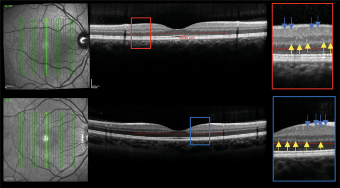
Background and objectives: Microglia, the resident immune cell of the brain and retina, is widespread activated in the white and gray matter (GM) in multiple sclerosis (MS). The objective of this study is to evaluate the presence and number of hyperreflecting foci (HRF), considered clusters of activated and proliferating retinal microglia, and their association with clinical and radiologic disease parameters in relapsing-remitting MS (RRMS).

Methods: At baseline, 80 patients with RRMS underwent optical coherence tomography (OCT) and 3T-MRI (including 3-dimensional T1, fluid-attenuated inversion recovery, and double inversion recovery sequences), closed to their disease onset (6.3 ± 5.1 months). These patients were then clinically and radiologically followed up for a mean of 43 months, evaluating the no evidence of disease activity (NEDA) condition, further divided into clinical (cNEDA) and radiologic (rNEDA). Patients with a clinical history or MRI/OCT findings suggestive of optic neuritis (ON) were excluded from the study.

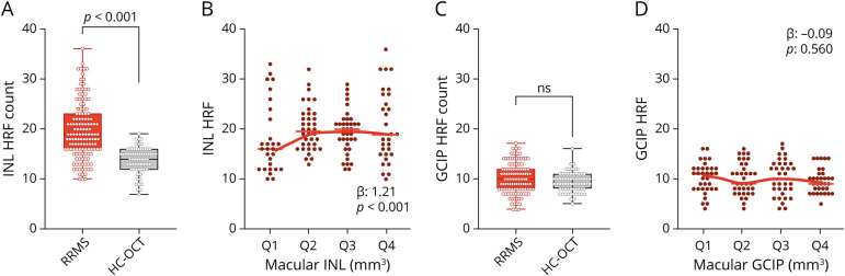
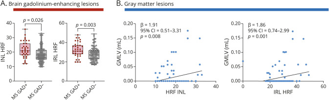
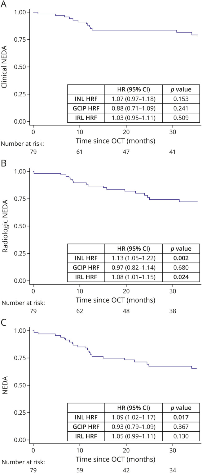
Results: Compared with healthy controls, the HRF number was significantly higher in the inner nuclear layer (INL) of patients with RRMS (19.55 ± 5.65 vs 13.84 ± 2.57, p < 0.001) and associated with INL volume (β: 1.21, p < 0.001). GM lesion volume significantly correlated with the INL HRF count (p = 0.008). Survival analysis revealed a significant association between INL HRF and both cNEDA (p = 0.017) and rNEDA (p = 0.002).

Discussion: We found a strong association between retinal microglial proliferation and cortical pathology in RRMS, a finding suggesting a possible underlying common immunopathologic mechanism. Furthermore, microglial activation at baseline was observed to predict subsequent inflammatory events, indicating that HRF might be a candidate prognostic biomarker worthy of further investigation.

Classification of evidence: This study provides Class II evidence that in patients with early RRMS but without ON, the number of HRF on OCT of the retinal inner nuclear layer is associated with cNEDA and rNEDA.


Figures

Figure 1
Figure 1. HRF Protocol
Macular scans and HRF visualization in RRMS (upper image) and HC (lower image); INL foci are indicated by yellow arrows and ganglion cell and inner plexiform layer (GCIP) HRF by blue arrows. HRF were counted in the area included between 2 perpendicular lines to Bruch membrane traced at 1,500 μm both temporally and nasally from the center of the fovea. HRF were defined as isolated, small size (<30 μm), punctiform elements with moderate reflectivity but without any back shadowing. HC = healthy control; HRF = hyperreflecting foci; INL = inner nuclear layer; RRMS = relapsing-remitting multiple sclerosis.
Figure 2
Figure 2. HRF Are Increased in the INL of Patients With RRMS and Associate With Macular INL Volume
(A) HRF were significantly increased in the INL of RRMS. (B) INL HRF count associated with INL volume (volumes were clustered in quartiles, from lowest values, Q1, to highest, Q4). (C) The HRF count in the GCIP did not differ between RRMS and HC and (D) was not associated with GCIP volume. Each dot represents 1 eye; however, the shown p values refer to the analysis with generalized estimating equation models (see Statistical Analysis in Methods). HC = healthy control; HRF = hyperreflecting foci; INL = inner nuclear layer; OCT = optical coherence tomography; RRMS = relapsing-remitting multiple sclerosis.
Figure 3
Figure 3. HRF Associated With Brain Inflammatory Parameters
(A) Both IRL and INL HRF count are increased in the presence of brain gadolinium-enhancing lesion. (B) Both INL and IRL HRF count associated with GMLV. Each dot represents 1 eye; however, the shown p values refer to the analysis with generalized estimating equation models (see Statistical Analysis in Methods). GMLV = gray matter lesion volume; HRF = hyperreflecting foci; INL = inner nuclear layer; IRL = inner retinal layer; MS = multiple sclerosis.
Figure 4
Figure 4. INL HRF Count Associated With NEDA Condition
(A) Clinical NEDA was not associated with the HRF count. (B) Radiologic NEDA associated with both INL and IRL HRF count. (C) NEDA condition associated with the INL HRF count. HRF = hyperreflecting foci; INL = inner nuclear layer; IRL = inner retinal layer; NEDA = no evidence of disease activity; OCT = optical coherence tomography.

References

    1. Colonna M, Butovsky O. Microglia function in the central nervous system during health and neurodegeneration. Annu Rev Immunol. 2017;35:441-468. doi:10.1146/annurev-immunol-051116-052358. - DOI - PMC - PubMed
    1. Spangenberg EE, Green KN. Inflammation in Alzheimer's disease: lessons learned from microglia-depletion models. Brain Behav Immun. 2017;61:1-11. doi:10.1016/j.bbi.2016.07.003. - DOI - PMC - PubMed
    1. Lucchinetti CF, Popescu BFG, Bunyan RF, et al. . Inflammatory cortical demyelination in early multiple sclerosis. N Engl J Med. 2011;365(23):2188-2197. doi:10.1056/nejmoa1100648. - DOI - PMC - PubMed
    1. Bø L, Vedeler CA, Nyland H, Trapp BD, Mørk SJ. Intracortical multiple sclerosis lesions are not associated with increased lymphocyte infiltration. Mult Scler. 2003;9(4):323-331. doi:10.1191/1352458503ms917oa. - DOI - PubMed
    1. van Horssen J, Singh S, van der Pol S, et al. . Clusters of activated microglia in normal-appearing white matter show signs of innate immune activation. J Neuroinflammation. 2012;9:156. doi:10.1186/1742-2094-9-156. - DOI - PMC - PubMed

Publication types

MeSH terms