Review
. 2022 May 23;10(1):40.
doi: 10.1038/s41413-022-00212-1.

Prospects and challenges of dynamic DNA nanostructures in biomedical applications

Affiliations
Review

Prospects and challenges of dynamic DNA nanostructures in biomedical applications

Taoran Tian et al. Bone Res. .

Abstract

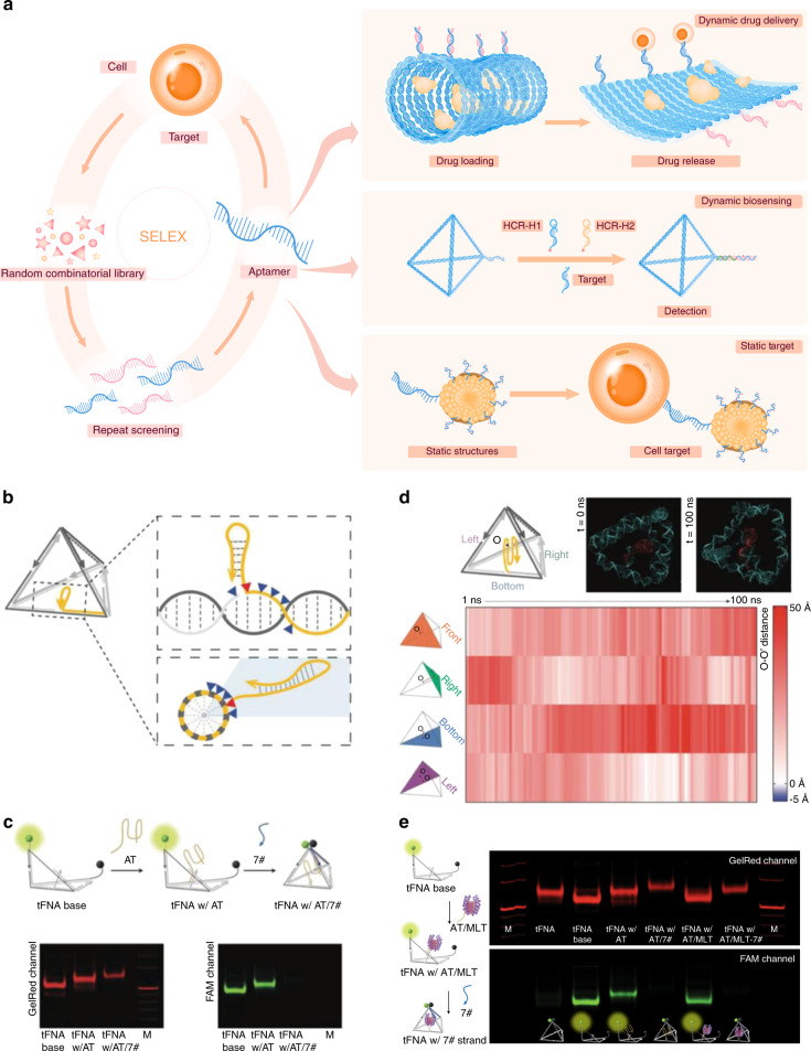
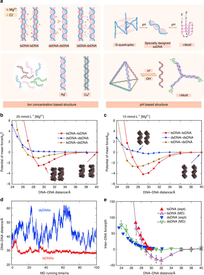
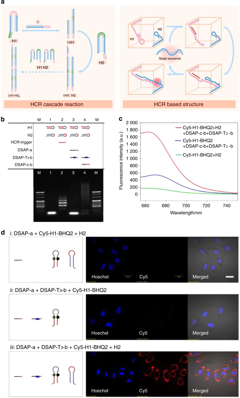
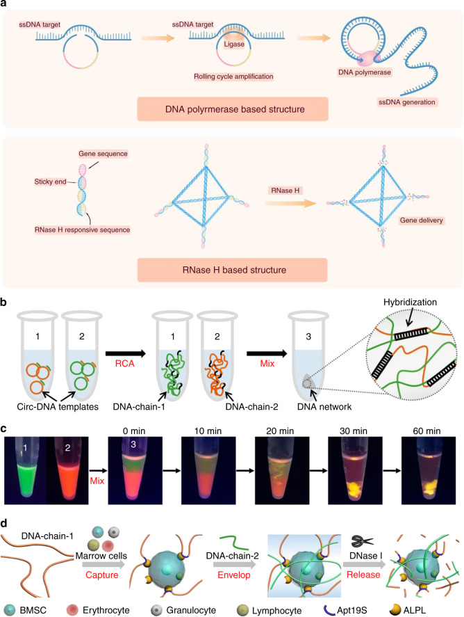
The physicochemical nature of DNA allows the assembly of highly predictable structures via several fabrication strategies, which have been applied to make breakthroughs in various fields. Moreover, DNA nanostructures are regarded as materials with excellent editability and biocompatibility for biomedical applications. The ongoing maintenance and release of new DNA structure design tools ease the work and make large and arbitrary DNA structures feasible for different applications. However, the nature of DNA nanostructures endows them with several stimulus-responsive mechanisms capable of responding to biomolecules, such as nucleic acids and proteins, as well as biophysical environmental parameters, such as temperature and pH. Via these mechanisms, stimulus-responsive dynamic DNA nanostructures have been applied in several biomedical settings, including basic research, active drug delivery, biosensor development, and tissue engineering. These applications have shown the versatility of dynamic DNA nanostructures, with unignorable merits that exceed those of their traditional counterparts, such as polymers and metal particles. However, there are stability, yield, exogenous DNA, and ethical considerations regarding their clinical translation. In this review, we first introduce the recent efforts and discoveries in DNA nanotechnology, highlighting the uses of dynamic DNA nanostructures in biomedical applications. Then, several dynamic DNA nanostructures are presented, and their typical biomedical applications, including their use as DNA aptamers, ion concentration/pH-sensitive DNA molecules, DNA nanostructures capable of strand displacement reactions, and protein-based dynamic DNA nanostructures, are discussed. Finally, the challenges regarding the biomedical applications of dynamic DNA nanostructures are discussed.


Conflict of interest statement

The authors declare no competing interests.

Figures

Fig. 1
Fig. 1
Dynamic DNA structures based on DNA aptamers. a Schematic illustration of the screening process for aptamer and aptamer-based nucleic acid nanostructures. b Continuous sites were screened for cargo loading via PAGE (screened sites are denoted as blue triangles). The best loading site (shown in the panel, red triangle) was selected by lagging tag modification (yellow loop at the 3′ end). c The cargo loading strand (attachment strand, AT strand) was loaded into the exoskeleton with stepped annealing: the green sphere represents the 3′ modification of FAM, the black sphere represents the 5′ modification of BHQ-1. The fluorescence changes and band shifts in the PAGE gel indicated successful fabrication of the AT strand-loaded tFNA exoskeleton. d All-atom MD simulation of the AT strand-loaded tFNA exoskeleton revealed that the AT strand was well encapsulated inside the tFNA exoskeleton. The distances between the center of mass of the G-quadruplex strand (O) and the four surfaces of the tFNA (O-O′) were calculated to be positive in the equilibrium state. e The MLT-loaded tFNA exoskeleton, the nanobee, was fabricated via three-step annealing and verified by PAGE gel electrophoresis. Copyright 2022, John Wiley and Sons
Fig. 2
Fig. 2
Dynamic DNA structures based on the ion concentration/pH. a Schematic illustration of dynamic DNA structures based on the ion concentration or pH. The mean force potentials between DNAs are given for three cases, namely, tsDNA-tsDNA, dsDNA-dsDNA, and tsDNA-dsDNA, in ∼20 (b) and ∼100 mmol·L−1 Mg2+ solutions (c). d Shown here is the distance between the mass center of the DNAs versus the MD simulation time from MD simulations for two tsDNAs (red) and two dsDNAs (blue) without the connection spring in a 20 mmol·L−1 Mg2+ solution. e The inter-DNA forces were calculated from our MD simulations and the experimental measurements on osmotic pressure. Copyright 2017, Biophysical Journal
Fig. 3
Fig. 3
Dynamic DNA structures based on strand displacement reactions. a Schematic illustration of dynamic DNA structures based on the strand displacement reaction. b The gel electrophoresis and c fluorescence characterization results of the HCR products induced by a DSAP using DSAP-c-b to simulate target recognition. Lane M: DNA marker. d Fluorescence confocal microscopy images of target SMMC-7721 cells after incubation with different DNA probes. (i) DSAP-a + Cy5-H1-BHQ2 + H2, (ii) DSAP-a + DSAP-T3-b + Cy5-H1-BHQ2, and (iii) DSAP-a + DSAP-T3-b + Cy5-H1-BHQ2 + H2. Cells were stained with Hoechst 33342 before imaging. (Scale bar: 20 μm). Copyright 2020, American Chemical Society
Fig. 4
Fig. 4
Dynamic DNA structures based on proteins or other substances. a Schematic illustration of dynamic DNA structures based on proteins. b Synthetic process of ultralong DNA chains via RCA to obtain a 3D DNA network. c Mix of DNA chains to visualize the molecular diffusion and phase inversion during the formation of a DNA network. DNA-chain-1 and DNA-chain-2 were stained with SYBR Green II and Gel Red, respectively. d The process of capture, envelopment, and release: (1) capture, DNA-chain-1 was incubated with BMSCs for cell capture by anchoring to the aptamer Apt19S; (2) envelopment, DNA-chain-2 was added to the cell-containing DNA-chain-1 solution to trigger the formation of the DNA network; and (3) release, the DNA network can be digested by the nuclease DNase I to release BMSCs. Copyright 2020, American Chemical Society
Fig. 5
Fig. 5
Assembly of nuclear pore complex mimics by DNA origami. a Schematic illustration of the fabrication of the DNA origami NPC mimic and its application in programmable protein arrangement. b As reported by Qi et al. two central channel nucleoporins of yeast origin, Nsp1 and Nup100, were expressed with a SNAP tag, which enables their conjugation with a benzylguanine-modified DNA oligo. c Illustration and TEM images of different protein-gated DNA origami NPC mimics. Scale bar = 50 nm. Copyright 2021, American Chemical Society
Fig. 6
Fig. 6
Drug transport and delivery with dynamic DNA nanostructures. The editability and physiochemical nature of DNA nanostructures allow various drug delivery strategies for dynamic and targeted delivery. a A repertoire of dynamic DNA nanostructures provides a stimulus-responsive lock and switch controlling cargo release. b DNA origami and DNA framework structures enable different cargo encapsulation strategies for cargo protection
Fig. 7
Fig. 7
DNA hairpin-endowed biosensors. a Schematic illustration of a DNA tetrahedron (TDN)-based biosensor (T-probe) and its enhanced imaging of the target via HCR. The red dot indicates the Cy5 module; the white dot indicates the BHQ module. b Agarose gel electrophoresis image of the fabrication and HCR assay. c Fluorescence intensity responses of the T-probe exposed to the target at different molecular ratios. d Comparison of the reaction distance between naked DNA hairpins and the T-probe. e The time-dependent intensity of the T-probe suggests that it is a rapid sensor platform. f and g Specificity and sensitivity evaluation of the T-probe system. Copyright 2022, John Wiley and Sons

Similar articles

Cited by

References

    1. Seeman NC. DNA in a material world. Nature. 2003;421:427–431. doi: 10.1038/nature01406. - DOI - PubMed
    1. Winfree E, Liu F, Wenzler LA, Seeman NC. Design and self-assembly of two-dimensional DNA crystals. Nature. 1998;394:539–544. doi: 10.1038/28998. - DOI - PubMed
    1. Seeman NC, Sleiman HF. DNA nanotechnology. Nat. Rev. Mater. 2017;3:17068. doi: 10.1038/natrevmats.2017.68. - DOI
    1. Pinheiro AV, Han D, Shih WM, Yan H. Challenges and opportunities for structural DNA nanotechnology. Nat. Nanotechnol. 2011;6:763–772. doi: 10.1038/nnano.2011.187. - DOI - PMC - PubMed
    1. Mathieu F, et al. Six-helix bundles designed from DNA. Nano Lett. 2005;5:661–665. doi: 10.1021/nl050084f. - DOI - PMC - PubMed