. 2022 May 23;13(1):2867.
doi: 10.1038/s41467-022-30464-8.

ADAMTS4-specific MR probe to assess aortic aneurysms in vivo using synthetic peptide libraries

Affiliations

ADAMTS4-specific MR probe to assess aortic aneurysms in vivo using synthetic peptide libraries

Jan O Kaufmann et al. Nat Commun. .

Abstract

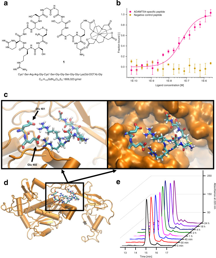
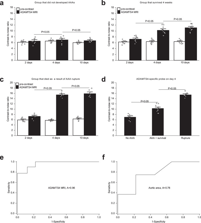
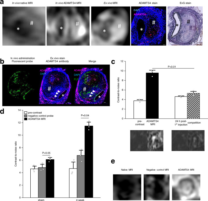
The incidence of abdominal aortic aneurysms (AAAs) has substantially increased during the last 20 years and their rupture remains the third most common cause of sudden death in the cardiovascular field after myocardial infarction and stroke. The only established clinical parameter to assess AAAs is based on the aneurysm size. Novel biomarkers are needed to improve the assessment of the risk of rupture. ADAMTS4 (A Disintegrin And Metalloproteinase with ThromboSpondin motifs 4) is a strongly upregulated proteoglycan cleaving enzyme in the unstable course of AAAs. In the screening of a one-bead-one-compound library against ADAMTS4, a low-molecular-weight cyclic peptide is discovered with favorable properties for in vivo molecular magnetic resonance imaging applications. After identification and characterization, it's potential is evaluated in an AAA mouse model. The ADAMTS4-specific probe enables the in vivo imaging-based prediction of aneurysm expansion and rupture.


Conflict of interest statement

The authors declare no competing interests.

Figures

Fig. 1
Fig. 1. Introduction of a one-bead-one-compound-library for screening against ADAMTS4 and evaluation of the binding properties of the discovered low-molecular-weight cyclic highly selective peptide targeted against ADAMTS4.
a A one-bead-one-compound-library was used to screen for binders against ADAMTS4. First, we adapted our on-chip screening technique to the requirements of in vivo MR imaging with an emphasis on cyclic peptides to achieve high peptide stability against proteases and high binding affinities. For the screening, we used our target protein, which was detected by an anti-His-Ab. The anti-His-Ab was afterward stained with an anti-Mouse IgG-Atto 633 antibody. After immobilization of the beads on a chip, firstly, a prescan against both antibodies was performed, to exclude positive hits against the antibodies. This was followed by the main scan against ADAMTS4 and a second scan against ADAMTS5. After each step, the chip was regenerated by guanidine hydrochloride. For MALDI-TOF-MS identification, the peptides were cleaved from the resin with ammonia gas, and a matrix was applied. In the last step, the amino acid sequence can be determined by MALDI-TOF-MS on-chip. b During the screening, different types of hits were detected; positive hits against (from left to right) the antibodies (cyan), against ADAMTS4 and ADAMTS5, only ADAMTS4 (magenta), or only ADAMTS5 (white). MALDI-TOF-MS: Matrix-assisted laser desorption/ionization-time of flights-mass spectrometry; MR: magnetic resonance.
Fig. 2
Fig. 2. Evaluation of the binding affinity (including negative control peptide), binding site, and stability of the ADAMTS4-specific probe.
a The ADAMTS4-specific probe contains 14 amino acids with a small cyclic ring of six amino acids. The MR active DOTA-Gd complex is coupled via a lysine side chain. The binding part and the MR active part are linked by a polar linker of glycine and serine amino acids. b By using MST, the binding constant between ADAMTS4 and the Cy5-labeled probe could be determined and showed a medium nanomolar affinity of 51 ± 21 nM, which is in the ideal range for specific MRI probes. However, for the negative control peptide, we could not find binding in this concentration range. As negative control, we used the peptide sequence from the screening, which showed also very low interaction in the SPR experiment with the sequence Cys*-Phe-His-Pro-Tyr-Cys*-Ser (n = 3 biologically independent measurements for each peptide; the data are presented as mean values ± SD). c Using molecular docking for predicting putative interaction geometries between a model structure of ADAMTS4 and the ADAMTS4-specific cyclic peptide, a binding cleft in ADAMTS4 was identified, in which the simultaneous contact between two glutamic acid residues from the protein and the two arginine residues of the peptide is indicated. Additionally, the peptide shows high contact to the ADAMTS4 protein (space-filled structure) and no dissociation during an MD simulation was observed. The binding cleft is unique for ADAMTS4 and was not found for ADAMTS1 and ADAMSTS5. d The binding cleft is located near to the protein surface and since the shortened linker of the peptide pointed outside, access to water is feasible, which is required for the generation of the T1 signal of the MR probe. e No relevant degradation of the ADAMTS4-specific probe within 24 h was measured in the human plasma by HPLC. HPLC High-performance liquid chromatography, MST Microscale thermophoresis, nM nanomolar, KD: binding constant; MRI magnetic resonance imaging. Source data for b, e are provided as a Source Data file.
Fig. 3
Fig. 3. In vivo MR imaging using a low-molecular-weight cyclic highly selective peptide targeted against ADAMTS4.
ac Time-of-flight angiograms show the suprarenal abdominal aorta of male apolipoprotein-E knockout mice (first row). a A sham-operated mouse receiving saline showed no pathological changes of the aortic wall in vivo and no ADAMTS4 expression. Two (b) and 4 weeks (c) following angiotensin-infusion, aortic dilatation (white arrows) and a clear signal enhancement in the aneurysmal wall were observed on T1-weighted sequences after administration of the ADAMTS4-specific probe (arrow-heads). Ex vivo histology confirmed the formation of abdominal aortic aneurysm and a strong expression of ADAMTS4 within the aneurysmal wall (arrow-heads). d A significant increase of contrast-to-noise-ratio was observed after administration of the ADAMTS4-specific probe on in vivo MRI scans with a significantly stronger enhancement between sham animals (n = 10), the 2 weeks group (n = 10) (P = 0.0001), and the 4 weeks group (n = 10) (P = 0.04). Data are presented as mean values ± SEM. e Mice of the longitudinal group (n = 12) that developed an extensive progression of the abdominal aneurysm after 4 weeks of angiotensin II—infusion showed a significantly higher MR signal enhancement (P = 0.0008) after injection of the ADAMTS4-specific probe compared to mice with no further aneurysm progression. Data are presented as mean values ± SEM. f In vivo contrast-to-noise ratio measurements after 4 weeks (n = 12) correlated well with histological analysis (y = 3.48x + 3.63; R² = 0.80; P = 1.1E-09). TOF Time-of-flight, MRI magnetic resonance imaging, EvG Elastica van Gieson staining, HE Hematoxylin-Eosin staining, AAA abdominal aortic aneurysm, ECM extracellular matrix. * indicates the aortic lumen; # indicates the thrombus area; Scale bars represent 100 µm; Source data for d, f are provided as a Source Data file. For data analysis, a two-sided, unpaired t-test was used.
Fig. 4
Fig. 4. Contrast-to-noise ratio for early detection of aneurysm and rupture prediction.
a The group that did not develop an AAA did not show a significant increase in ADAMTS4-MRI levels 2, 4, or 10 days after the initiation of AngII-infusion (n = 8, P = 0.21; P = 0,57). Data are presented as mean values ± SEM. b The group (n = 12) that developed an AAA however survived for 4 weeks showed a moderate increase of ADAMTS4-MRI levels after 4 days (P = 9.1E-06) with no further significant increase after 10 days (P = 0.14). Data are presented as mean values ± SEM. c The fatal AAA rupture group (n = 8) showed the strongest increase in ADAMTS4-MRI levels of the 4 days (P = 1.79E-06), with no further significant increase after 10 days (P = 0.19). Data are presented as mean values ± SEM. d ADAMTS4-MRI levels measured after 4 days significantly increased in the group that developed in AAA and survived for 4 weeks, compared to the group that did not develop any AAA (P = 0.001). The group that suffered a fatal AAA rupture showed further significant in increase ADAMTS4-MRI levels at this time point (P = 3.23E-05). The 4-day time point therefore seems to be the most promising time point for the prediction of an AAA development and survival. Data are presented as mean values ± SEM. e The ROC analysis demonstrates that the ADAMTS4-MRI level measured after 4 days enables of the prediction of a fatal AAA rupture with a sensitivity of 0.88 and a specificity of 0.9 (ROC Curve Area 0.96, Standard Error 0.04, 95% Confidence Interval 0.90 To 1.04). f The ROC analysis demonstrates that the aortic area measured after 4 days enables of the prediction of a fatal AAA rupture with a sensitivity of 0.78 and a specificity of 0.76 (ROC Curve Area 0.78, Standard Error 0.11, 95% Confidence Interval 0.55–1.01). AAA abdominal aortic aneurysm, MRI magnetic resonance imaging, AngII angiotensin II, ROC receiver operator characteristic Source data for af are provided as a Source Data file. For data analysis, a two-sided, unpaired t-test was used.
Fig. 5
Fig. 5. Confirmation of the specific binding of the ADAMTS4-specific probe.
a From left to right: In vivo MRI before and after administration of the ADAMTS4-specific probe in one male mouse 4 weeks following aneurysms induction was highly comparable to corresponding ex vivo MRI scan of the abdominal aorta and ex vivo immunofluorescence staining (ADAMTS4 stain) and Elastica van Gieson (EvG) staining. b Intravenous administration of the fluorescent variant of the ADAMTS4-specific probe resulted in a strong fluorescence of the aneurysmal wall that was clearly co-localized with areas of ADAMTS4 expression using immunofluorescence staining (white arrows). c The competition experiment (n = 3 mice) showed a significant decrease in the contrast-to-noise-ratio compared to the administration of the gadolinium-labeled ADAMTS4-specific probe alone, confirming the specific binding of the probe (P = 0.0025). Data are presented as mean values ± SEM. d, e Using the negative control probe Cys*-Phe-His-Pro-Tyr-Cys*-linker (DOTA-Gd) with a similar size to the ADAMTS4-specific probe, no significant increase in contrast-to-noise ratio (CNR) was measured in vivo in the aortic wall of ApoE-/- mice (n = 6). The application of the ADAMTS4-specific probe resulted in a strong and significant increase in CNR in the vessel wall as well in the thrombus. *P = 0.08; **P = 0.04; Data are presented as mean values ± SEM. FLP Fluorescein-labeled probe, Scale bars indicate 100 µm. * indicate the aortic lumen; # indicates the thrombus area. Source data for c, d are provided as a Source Data file. For data analysis, a two-sided, unpaired t-test was used.
Fig. 6
Fig. 6. Ex vivo expression of ADAMTS4.
a Immunofluorescence analysis showed a significant increase in ADAMTS4-expression 2 and 4 weeks after aneurysm induction (n = 10 per group) (*P = 9.35E-07; **P = 6.31E-05). Data are presented as mean values ± SEM. b In vivo contrast-to-noise-ratio (CNR) measurements were in good correlation with ex vivo measurements of ADAMTS4 expression using immunofluorescence (n = 28; y = 0.95x + 7.73; R2 = 0.75, P = 4.08E-15). c Immunofluorescence staining showed a strong expression of ADAMTS4 and the associated CD68 macrophages within the aneurysmal wall after two and 4 weeks as well as a clear colocalization between areas of macrophage accumulation and ADAMTS4 expression. Sham-operated mice showed no relevant expression of CD68 or ADAMTS4. Scale bars represent 100 µm. d CD68- and ADAMTS4-expression within the aortic wall correlated well, using immunofluorescence analysis (n = 16; y = 1.17x + 0.90; R2 = 0.80, P = 0.001). e, f Example of a ruptured human aortic aneurysm, a colocalization of the ADAMTS4 (e) with macrophages (f) can be appreciated. Sections were incubated with polyclonal ADAMTS4 antibody (Rabbit anti-mouse ADAMTS4) and monoclonal CD68 antibody (Rat anti-mouse CD68). Source data for a, b, d are provided as a Source Data file. For data analysis, a two-sided, unpaired t-test was used.
Fig. 7
Fig. 7. Correlation of in vivo MRI with inductively coupled plasma mass spectrometry (ICP-MS) and spatial localization of Gd using laser-ablation inductively coupled plasma mass spectrometry (LA-ICP-MS).
a Inductively coupled plasma mass spectrometry (ICP-MS) analysis showed a good correlation between contrast-to-noise-ratio measurements and Gd amount in the aortic wall (n = 7; y = 0.10x + 6.77; R2 = 0.57, P = 0.02). b First row: Laser-ablation inductively coupled plasma mass spectrometry visualized the gadolinium-based ADAMTS4-specific probe within the aortic wall (black arrowheads) of a mouse 4 weeks after aneurysm induction (left hand) and of a sham-operated mouse (right hand) by the Gd-signal. Second row: A clear colocalization of gadolinium-accumulation with areas positive for ADAMTS4 using immunofluorescence was observed (white arrowheads). Scale bars indicate 100 µm. * indicate the aortic lumen; # indicates the thrombus area. Source data for a are provided as a Source Data file. For data analysis, a two-sided, unpaired t-test was used.

References

    1. Roth GA, et al. Global burden of cardiovascular diseases and risk factors, 1990-2019: update from the GBD 2019 study. J. Am. Coll. Cardiol. 2020;76:2982–3021. doi: 10.1016/j.jacc.2020.11.010. - DOI - PMC - PubMed
    1. Gillum RF. Epidemiology of aortic aneurysm in the United States. J. Clin. Epidemiol. 1995;48:1289–1298. doi: 10.1016/0895-4356(95)00045-3. - DOI - PubMed
    1. Bengtsson H, Sonesson B, Bergqvist D. Incidence and prevalence of abdominal aortic aneurysms, estimated by necropsy studies and population screening by ultrasound. Ann. N. Y. Acad. Sci. 1996;800:1–24. doi: 10.1111/j.1749-6632.1996.tb33294.x. - DOI - PubMed
    1. Weiss N, Rodionov RN, Mahlmann A. Medical management of abdominal aortic aneurysms. Vasa. 2014;43:415–421. doi: 10.1024/0301-1526/a000388. - DOI - PubMed
    1. Johnston KW, et al. Suggested standards for reporting on arterial aneurysms. Subcommittee on reporting standards for arterial aneurysms, Ad Hoc committee on reporting standards, society for vascular surgery and north American chapter, International society for cardiovascular surgery. J. Vasc. Surg. 1991;13:452–458. doi: 10.1067/mva.1991.26737. - DOI - PubMed

Publication types

Substances