. 2022 May 23;13(1):2861.
doi: 10.1038/s41467-022-30311-w.

Synthetic lethality between TP53 and ENDOD1

Affiliations

Synthetic lethality between TP53 and ENDOD1

Zizhi Tang et al. Nat Commun. .

Abstract

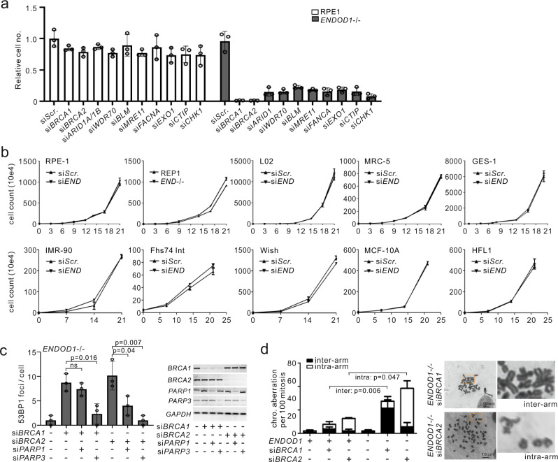
The atypical nuclease ENDOD1 functions with cGAS-STING in innate immunity. Here we identify a previously uncharacterized ENDOD1 function in DNA repair. ENDOD1 is enriched in the nucleus following H2O2 treatment and ENDOD1-/- cells show increased PARP chromatin-association. Loss of ENDOD1 function is synthetic lethal with homologous recombination defects, with affected cells accumulating DNA double strand breaks. Remarkably, we also uncover an additional synthetic lethality between ENDOD1 and p53. ENDOD1 depletion in TP53 mutated tumour cells, or p53 depletion in ENDOD1-/- cells, results in rapid single stranded DNA accumulation and cell death. Because TP53 is mutated in ~50% of tumours, ENDOD1 has potential as a wide-spectrum target for synthetic lethal treatments. To support this we demonstrate that systemic knockdown of mouse EndoD1 is well tolerated and whole-animal siRNA against human ENDOD1 restrains TP53 mutated tumour progression in xenograft models. These data identify ENDOD1 as a potential cancer-specific target for SL drug discovery.


Conflict of interest statement

A patent application ‘Application of reagents or drugs inhibiting an endonuclease in cancer therapy (202010295776.6)’ was filed on April 15, 2020.

Figures

Fig. 1
Fig. 1. Characterization of ENDOD1 in DNA repair.
a Schematic of ENDOD1 protein showing the predicted signal peptide (residues 1–22), the endonuclease domain (residues 49–257) and three C-terminal transmembrane motifs (Ts). b Left: Representative indirect immunofluorescence images for α-ENDOD1 (ABclonal) and α-PAR: untreated RPE1 cells (top row), RPE1 and ENDOD1−/− cells after a 5 min treatment with 10 mM H2O2 (bottom two rows). For the merged panels nuclear DNA was counterstained with DAPI. Right: quantification for nuclear signals at the indicated time points after H2O2 treatment. arb. units: arbitrary units. n = 3–5 biologically independent experiments. c Comet assay to assess repair efficiency. Left: quantification of tail moments from three repeats for proliferating RPE1 and ENDOD1−/− cells at the indicated time points after H2O2 challenge (p = 0.197 for RPE 35v ENDOD1−/− 35, Kruskal test). Middle: tail moments from 150 cells from each of 3 biologically independent experiments for serum-starved G1 arrested RPE1 and ENDOD1−/− cells at indicated time points after H2O2 challenge (p = 0.787 for RPE1 20 v ENDOD1−/− 20; p = 0.0000159 for RPE1 35v ENDOD1−/− 35, Kruskal test). Right: representative images of alkaline comet assay. Percentage tail moment was calculated by dividing the pixel intensity of tails by that of heads. White dot: median. Thick whisker: third quartile. Thin whisker: upper/lower adjacent values (1.5x inter-quartile range). d Relative viability of RPE1 or ENDOD1−/− cells treated with two different siPARP1 (si001 and si002) 48 h before challenge, with or without continuous H2O2 treatment (10 μM). Assay: CCK8 colorimetry. Inset showing the knockdown efficiency of each siRNA. n = 3 biologically independent experiments. Significance test: two-tailed Student’s t test. p values: 0.0057, 0.047. ns: not significant. e. Whole-cell extract (total), nuclear extracts (P1) and MNase-digested extract (fraction D) from RPE1 or ENDOD1−/− cells probed for PARP1, PARP2 and PARP3. Cells were treated with 0.01% MMS for 30 min. Histone H3 serves as a control. Representative image of 3 independent experiments.
Fig. 2
Fig. 2. Depletion of ENDOD1 causes SL with HRD.
a Relative viability of ENDOD1−/− and control RPE1 cells subjected to the indicated siRNA treatments. siRNA was transfected three times with three-day intervals. Cell viability was determined by CCK8 colorimetric assay. n = 3 biologically independent experiments. b Proliferation was quantified by haemocytometer cell counting over 3 weeks for the indicated immortalized non-cancerous cells with control siRNA (siScr.) or siENDOD1 treatment every 3 days. Control experiments using RPE1 and ENDOD1−/− are shown in the first two panels. c Left: quantification of 53BP1 foci in ENDOD1−/− cells 72 h following treatment with the indicated siRNAs. n = 3 biologically independent experiments. Error bars: SEM. Significance test: two-tailed Student’s t test. ns: not significant. Right: semi-quantitative PCR showing knockdown efficiency. d. Ablation of BRCA1 or BRCA2 in ENDOD1 null cells results in increased chromosome aberrations. Left: quantification of inter- and intra-chromosomal aberrations in RPE1 or ENDOD1−/− cells treated with siBRCA1 or siBRCA2. n = 3 biologically independent experiments. Error bars: SEM. Significance test: two-tailed Student’s t test. Right; representative images.
Fig. 3
Fig. 3. Concomitant loss of ENDOD1 and p53 causes SL.
a Proliferation curves for representative cancer cell lines following either siENDOD1 or control transfection. n = 3 biologically independent experiments. Error bars: SEM. At 3-day intervals cells were passaged, counted by haemocytometer and transfected. Reported TP53 status is indicated. b. Correlation of TP53/HR mutations with cytotoxicity of siENDOD1 in terms of inhibition rate (%) for cancer cell lines (each point represents mean of n = 3 biologically independent experiments). Error bars; SEM. c Relative viability determined by CCK8 colorimetry of ENDOD1−/− and control RPE1 cells 5 and 7 days after treatment with two different siTP53 (si001 and si002). n = 3 biologically independent experiments. Error bars: SEM d Equivalent experiment as in (c), using cells arrested in G1 by serum starvation. n = 3 biologically independent experiments. Error bars: SEM. All significance tests: two-tailed Student’s t test.
Fig. 4
Fig. 4. SL between ENDOD1 and TP53 correlates with ssDNA formation.
a Quantification of tail moments for neutral (bottom) and alkali (top) comet assays in G1-arrested ENDOD1−/− and control RPE1 cells treated with siTP53 or control siRNA. n = 150 cells from each of 3 biologically independent experiments. White dot: median. Thick whisker: third quartile. Thin whisker: upper/lower adjacent values (1.5x inter-quartile range). b Immunofluorescent staining for pRPA32 in ENDOD1−/− and control RPE1 G1 arrested cells 96 h following siTP53 or control treatment. Merged image is with DAPI staining. c Equivalent experiment as in (b), non-denatured staining for BrdU that was incorporated into cells before serum starvation. d Non-denatured BrdU signals in ENDOD1−/− siTP53 cells with or without prior S1 nuclease digestion. Representative of 3 independent experiments. e Quantification of agarose gel band intensity of undigested genomic DNA purified from the indicated cells after treatment with the specified units of S1 nuclease. Representative image of 3 independent experiments. A representative original gel for 0.02 U/μl is shown in Supplementary Fig. 5e. f pRPA32 staining in cancer cell lines that harbour TP53 mutations 72 h after treatment with control (siScr.) or siENDOD1. A549 is a TP53 wild type control. g. Quantification of pRPA32 foci in siENDOD1 treated SKOV-3 cells complemented with the indicated TP53 alleles. n = 3–4 biologically independent experiments. Error bars; SEM. Significance test: two-tailed Student’s t test.
Fig. 5
Fig. 5. PARP activity is required for the SL between ENDOD1 and TP53.
a pRPA32 foci in serum starved ENDOD1−/− cells treated with the specified siRNAs. n = 6 biologically independent experiments. Error bars: SEM. b. Immunoblotting for PARP1 on chromatin (fraction D—see Fig. 1e) in ENDOD1−/− and control RPE1 cells following siTP53 or siRNA control treatment. Representative image of 4 independent experiments. c Quantification of nuclear PAR staining 72 h following the indicated siRNA treatments of ENDOD1−/− and control RPE1 cells (left: cycling, middle: G1 arrested, right: immunoblot showing knockdown efficiency for siTP53). arb. units: arbitrary units. n = 381–731 cells. Red bar: median. Whiskers: SEM. d pRPA32 and PAR co-staining 72 h following the indicated siRNA treatments of ENDOD1−/− and control RPE1 cells. Representative image of 3 independent experiments. e. Quantification of PAR foci in SKOV-3 cells upon exogenous expression of the indicated TP53 alleles. arb. units: arbitrary units. n = 3–4 biologically independent experiments. f pRPA32 foci 3 days after serum starved ENDOD1−/− cells were treated with siTP53 and PARPi (10 nM Talazoparib, 100 nM Olaparib). n = 3 biologically independent experiments. Error bars: SEM. g Relative proliferation 6 days after ENDOD1−/− and control RPE1 cells were treated with siTP53 and PARPi (2 nM Talazoparib, 20 nM Olaparib) n = 3 biologically independent experiments. Error bars: SEM. h Talazoparib addition-removal assay in ENDOD1−/− cells. 10 nM Talazoparib was added upon siTP53 transfection and removed 72 h later (time 0). Cells were fixed at the indicated time for α-pRPA32 or α-PAR staining. Immunoblot evaluation for the knockdown efficiency of TP53 is shown on the right. arb. units: arbitrary units. n = 3 biologically independent experiments. Error bars; SEM. All significance tests: two-tailed Student’s t test.
Fig. 6
Fig. 6. SSBR and HR machinery provides key signals for ssDNA production.
a Quantification of pRPA32 foci in a Talazoparib addition-removal assay with siTP53 treated ENDOD1−/− cells co-treated with the indicated SSBR siRNAs. 10 nM Talazoparib was added upon siRNA treatment and removed 72 h later (time 0). n = 3 biologically independent samples. Error bars: SEM. b Quantification of a-PAR foci in G1 serum starved ENDOD1−/− and control RPE1 cells following treatment with the indicated siRNAs. n = (99-103) × 5 cells (each data point represents the average foci number of 5 cell counts). Red bar: median. Whiskers: SEM. c Quantification of XRCC1 foci in serum-starved ENDOD1−/− and control RPE1 cells following siTP53 treatment with or without co-treatment with Talazoparib. n = (99–100) × 5 cells (each data point represents the average foci number of 5 cell counts). Red bar: median. Whiskers: SEM. d pRPA32 and a-PAR foci in siTP53 treated ENDOD1−/− cells co-treated with and the indicated siRNAs targeting DNA end processing factors. n = 151–167 cells. e Quantification of CTIP, FANCD2 and MRE11 foci in a Talazoparib addition-removal assay with siTP53 treated ENDOD1−/− cells. n = 3 biologically independent experiments. Error bars: SEM. 10 nM Talazoparib was added upon siRNA treatment and removed 72 h later (time 0). f Schematic model for how ENDOD1 protects genomic integrity together with HR (left) and p53 (right). See discussion for details.
Fig. 7
Fig. 7. mEndod1 systemic knockdown is well tolerated.
a Body weight tracked through 90 days for the indicated in vivo knockdown groups (two injections per week). simWdr70 mice were sacrificed at 60 days due to severe disease. b Representative images of hematoxylin and eosin staining of paraffin-embedded sections from the indicated tissues. c Haemocytometer counts for peripheral blood cells at the endpoint of the experiment for each knockdown group. Control 90 days (n = 6 animals), simEndod1 90 days (n = 5 animals), simWdr70 60 days (n = 2 animals). Significance test: two-tailed Student’s t test. ns: not significant. d FACS analysis for peripheral myeloid and lymphoid cells when experiments terminated. n = 6, 5 and 2 animals for simScr, simEnd and simWdr70, respectively. Error bars: SEM. Significance test: two-tailed Student’s t test. ns: not significant. Cell surface markers used are shown in parentheses. e Equivalent FACS analysis as above for bone marrow HSC. f Anti-tumour treatment using whole animal in vivo knockdown of ENDOD1 for p53-deficient (SKOV-3 and C33A) xenograft models in nude mice. Volumes of individual tumours were measured. x-axis: treatment days. Numbers of animals (n) are indicated.

References

    1. Gelmon KA, et al. Olaparib in patients with recurrent high-grade serous or poorly differentiated ovarian carcinoma or triple-negative breast cancer: a phase 2, multicentre, open-label, non-randomised study. Lancet Oncol. 2011;12:852–861. doi: 10.1016/S1470-2045(11)70214-5. - DOI - PubMed
    1. Shen Y, et al. BMN 673, a novel and highly potent PARP1/2 inhibitor for the treatment of human cancers with DNA repair deficiency. Clin. Cancer Res. 2013;19:5003–5015. doi: 10.1158/1078-0432.CCR-13-1391. - DOI - PMC - PubMed
    1. Bryant HE, et al. Specific killing of BRCA2-deficient tumours with inhibitors of poly(ADP-ribose) polymerase. Nature. 2005;434:913–917. doi: 10.1038/nature03443. - DOI - PubMed
    1. Farmer H, et al. Targeting the DNA repair defect in BRCA mutant cells as a therapeutic strategy. Nature. 2005;434:917–921. doi: 10.1038/nature03445. - DOI - PubMed
    1. Helleday T. Putting poly (ADP-ribose) polymerase and other DNA repair inhibitors into clinical practice. Curr. Opin. Oncol. 2013;25:609–614. doi: 10.1097/CCO.0000000000000016. - DOI - PubMed

Publication types

LinkOut - more resources