. 2022 May 23;12(1):8682.
doi: 10.1038/s41598-022-10335-4.

A Fetal Brain magnetic resonance Acquisition Numerical phantom (FaBiAN)

Affiliations

A Fetal Brain magnetic resonance Acquisition Numerical phantom (FaBiAN)

Hélène Lajous et al. Sci Rep. .

Abstract

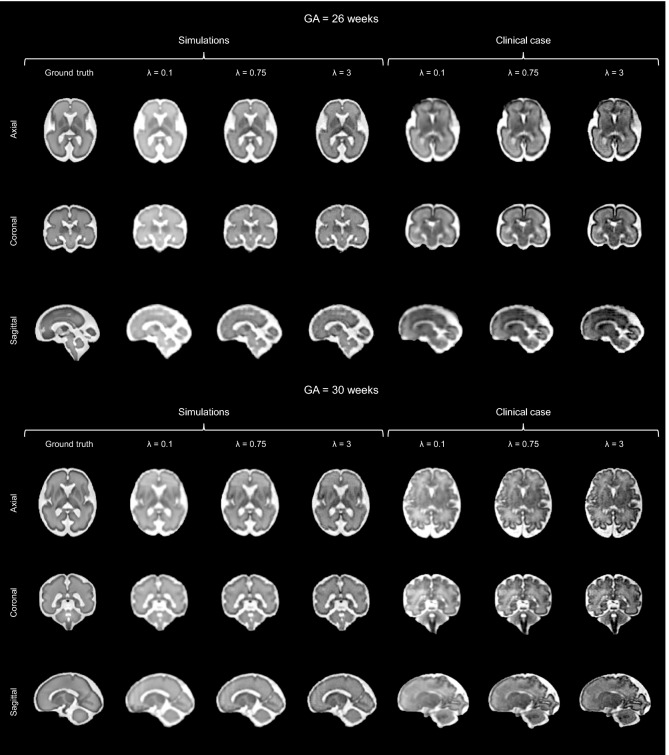
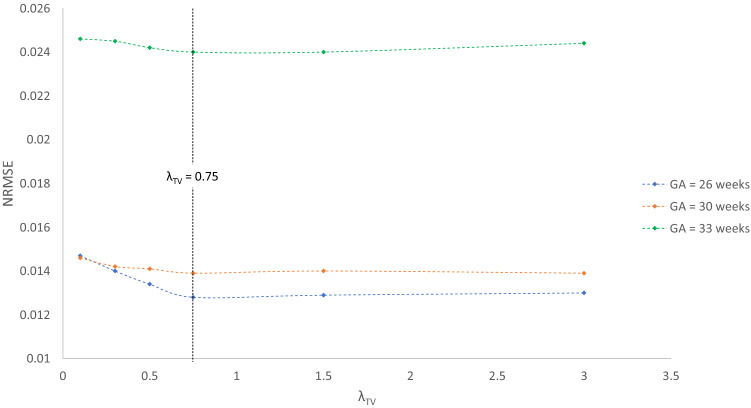
Accurate characterization of in utero human brain maturation is critical as it involves complex and interconnected structural and functional processes that may influence health later in life. Magnetic resonance imaging is a powerful tool to investigate equivocal neurological patterns during fetal development. However, the number of acquisitions of satisfactory quality available in this cohort of sensitive subjects remains scarce, thus hindering the validation of advanced image processing techniques. Numerical phantoms can mitigate these limitations by providing a controlled environment with a known ground truth. In this work, we present FaBiAN, an open-source Fetal Brain magnetic resonance Acquisition Numerical phantom that simulates clinical T2-weighted fast spin echo sequences of the fetal brain. This unique tool is based on a general, flexible and realistic setup that includes stochastic fetal movements, thus providing images of the fetal brain throughout maturation comparable to clinical acquisitions. We demonstrate its value to evaluate the robustness and optimize the accuracy of an algorithm for super-resolution fetal brain magnetic resonance imaging from simulated motion-corrupted 2D low-resolution series compared to a synthetic high-resolution reference volume. We also show that the images generated can complement clinical datasets to support data-intensive deep learning methods for fetal brain tissue segmentation.


Conflict of interest statement

The authors declare no competing interests.

Figures

Figure 1
Figure 1
Workflow for simulating images of the fetal brain acquired by a fast spin echo (FSE) sequence (i) from segmented HR anatomical MR images, illustrated for a fetus of 30 weeks of GA. (ii) Brain tissues are classified into gray matter, white matter and cerebrospinal fluid. (iii) Anatomical structures are converted to the corresponding MR contrast to obtain reference T1 and T2 maps of the fetal brain at either 1.5 or 3 T. (iv) The EPG algorithm allows to accurately simulate the T2 decay over time in every brain voxel by accounting for the effects of the stimulated echoes, as highlighted by the enlargement of the beginning of the curve. This spatiotemporal information is subsequently used (v) to sample the Fourier domain of the simulated images of the moving fetus. After the addition of noise to match the SNR of real clinical acquisitions, (vi) FSE images of the fetal brain are eventually recovered by 2D inverse Fourier transform (2D FT-1).
Figure 2
Figure 2
Visual inspection and comparison between clinical MR acquisitions and representative simulated HASTE images of the fetal brain in the three orthogonal orientations at four different GA (23, 26, 30 and 32 weeks). The amplitude of movement of the fetus is indicated from the motion index computation. Red arrows point out typical out-of-plane motion patterns.
Figure 3
Figure 3
Appreciation of the quality of SR reconstruction depending on the weight λ that controls the strength of the TV regularization. The potential of our framework FaBiAN for optimizing the reconstruction quality through parameter fine-tuning in the presence of motion is illustrated at two GA: 26 and 30 weeks. Two representative clinical cases are provided for comparison. The results for three values of λ are presented. For λ=0.1, the SR reconstruction looks blurry with poor tissue contrast. Using λ=3 improves the contrast but the images look noisy. For λ=0.75, the SR reconstruction is sharp with a contrast between different brain tissues similar to that observed in the 3D isotropic ground truth. Clinical cases from which the simulated HASTE images are derived highlight the accuracy of a SR reconstruction for this intermediate value of λ, especially with regards to the definition of the corpus callosum and the delineation of the cortex.
Figure 4
Figure 4
Normalized root mean squared error (NRMSE) between SR reconstructions from simulated data at a GA of 26, 30 and 33 weeks respectively and the corresponding 3D HR ground truth depending on the weight λ of the TV regularization. Six values of λ are tested: 0.1, 0.3, 0.5, 0.75, 1.5 and 3. The NRMSE is minimal for λ = 0.75.
Figure 5
Figure 5
(a) Normalized root mean squared error (NRMSE) and (b) mean structural similarity index (MSSIM) between SR reconstructions from different numbers of orthogonal LR HASTE series simulated at a GA of 30 weeks and the corresponding static 3D HR ground truth. The left panel shows results for motion-free data with various noise levels, a SD of 0.15 leading to a similar appearance as in clinical acquisitions. The right panel illustrates how the algorithm performs depending on the amplitude of fetal movements in the input series.
Figure 6
Figure 6
Appreciation of sharpness and tissue contrast enhancement in SR reconstructions from higher numbers of simulated orthogonal LR HASTE images corrupted by little motion at a GA of 30 weeks in comparison with the corresponding static 3D HR ground truth. The frontal cortex looks smoother and the putamen area sharper in the SR reconstruction from nine series compared to the SR reconstruction from three series. The mapping of local SSIM values and the computation of the MSSIM over the corresponding region-of-interest further support these observations.
Figure 7
Figure 7
Illustration of the accuracy of fetal brain tissue segmentation in a subject of 30.6 weeks of GA on (a) an axial slice from the SR reconstruction. Comparison of (b) the reference manual annotations, (c) the segmentation results obtained when performing extensive standard data augmentation on the clinical SR reconstructions (configuration (E), C15/S0), (d) the segmentation results obtained by the configuration (D) that complements this original dataset with fifteen additional simulated subjects (C15/S15), overlaid on the SR image. The segmentation of the cortex especially looks more accurate in (d), with an increased sensitivity to folding as highlighted by the white arrows.

References

    1. Volpe JJ. Brain injury in premature infants: A complex amalgam of destructive and developmental disturbances. Lancet Neurol. 2009;8:110–124. doi: 10.1016/S1474-4422(08)70294-1. - DOI - PMC - PubMed
    1. Ramírez-Vélez R. In utero fetal programming and its impact on health in adulthood. Endocrinol. Nutr. (English Ed.) 2012;59:383–393. doi: 10.1016/j.endonu.2012.02.002. - DOI - PubMed
    1. Bhat S, Acharya UR, Adeli H, Bairy GM, Adeli A. Autism: Cause factors, early diagnosis and therapies. Rev. Neurosci. 2014;25:841–850. doi: 10.1515/revneuro-2014-0056. - DOI - PubMed
    1. Miller SL, et al. Antenatal antioxidant treatment with melatonin to decrease newborn neurodevelopmental deficits and brain injury caused by fetal growth restriction. J. Pineal Res. 2014;56:283–294. doi: 10.1111/jpi.12121. - DOI - PubMed
    1. Kwon EJ, Kim YJ. What is fetal programming?: A lifetime health is under the control of in utero health. Obstetr. Gynecol. Sci. 2017;60:506–519. doi: 10.5468/ogs.2017.60.6.506. - DOI - PMC - PubMed

Publication types

MeSH terms