. 2022 Nov;29(11):2288-2302.
doi: 10.1038/s41418-022-01016-w. Epub 2022 May 23.

Epithelial de-differentiation triggered by co-ordinate epigenetic inactivation of the EHF and CDX1 transcription factors drives colorectal cancer progression

Affiliations

Epithelial de-differentiation triggered by co-ordinate epigenetic inactivation of the EHF and CDX1 transcription factors drives colorectal cancer progression

Ian Y Luk et al. Cell Death Differ. 2022 Nov.

Abstract

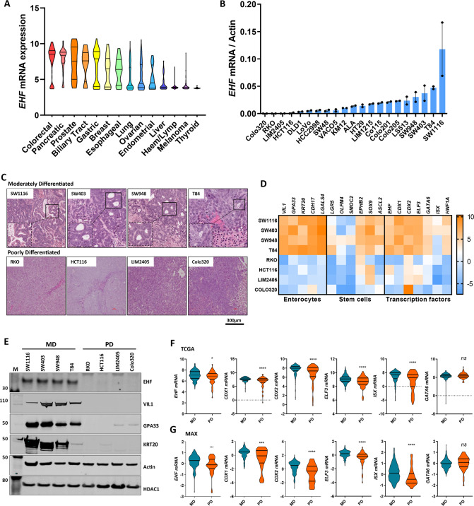
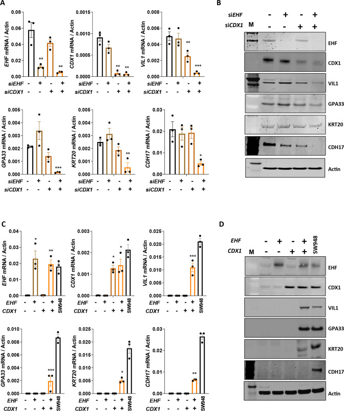
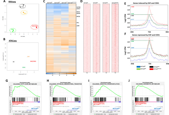
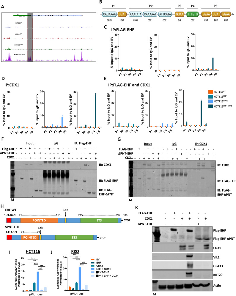
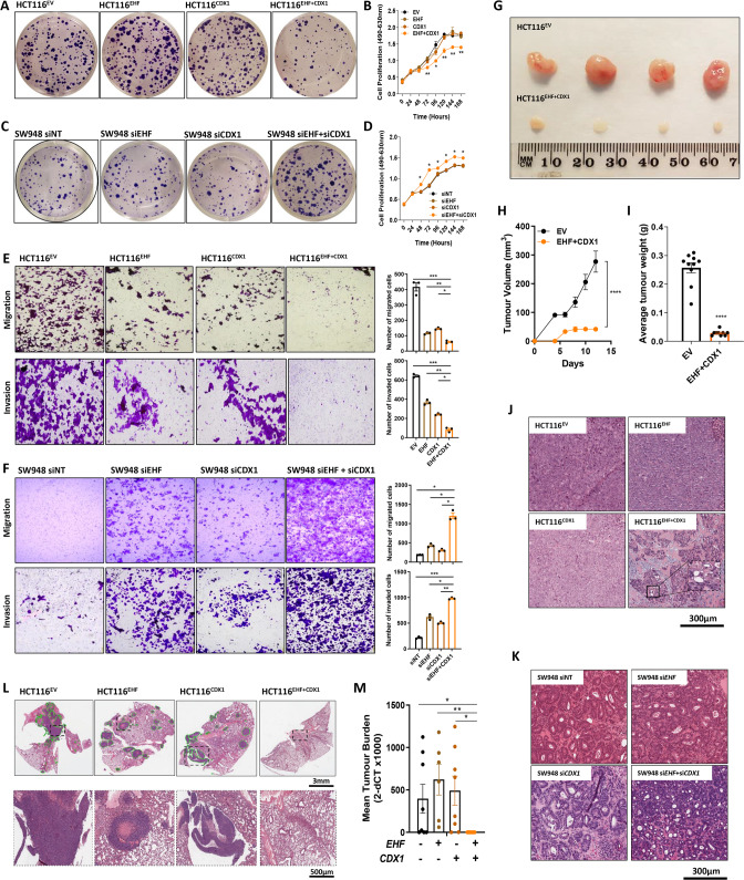
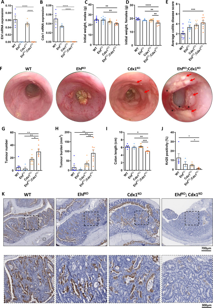
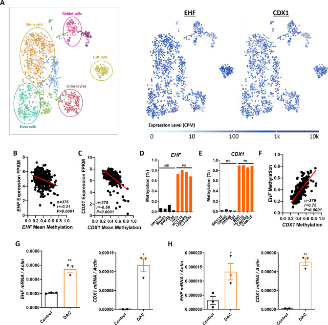
Colorectal cancers (CRCs) often display histological features indicative of aberrant differentiation but the molecular underpinnings of this trait and whether it directly drives disease progression is unclear. Here, we identify co-ordinate epigenetic inactivation of two epithelial-specific transcription factors, EHF and CDX1, as a mechanism driving differentiation loss in CRCs. Re-expression of EHF and CDX1 in poorly-differentiated CRC cells induced extensive chromatin remodelling, transcriptional re-programming, and differentiation along the enterocytic lineage, leading to reduced growth and metastasis. Strikingly, EHF and CDX1 were also able to reprogramme non-colonic epithelial cells to express colonic differentiation markers. By contrast, inactivation of EHF and CDX1 in well-differentiated CRC cells triggered tumour de-differentiation. Mechanistically, we demonstrate that EHF physically interacts with CDX1 via its PNT domain, and that these transcription factors co-operatively drive transcription of the colonic differentiation marker, VIL1. Compound genetic deletion of Ehf and Cdx1 in the mouse colon disrupted normal colonic differentiation and significantly enhanced colorectal tumour progression. These findings thus reveal a novel mechanism driving epithelial de-differentiation and tumour progression in CRC.


Conflict of interest statement

The authors declare no competing interests.

Figures

Fig. 1
Fig. 1. EHF expression is downregulated in poorly-differentiated colorectal cancers.
A EHF mRNA expression in 1080 cancer cell lines derived from various tumour types obtained from the Broad Institute Cancer Cell Line Encyclopaedia (CCLE) database. B Validation of EHF mRNA expression in 20 CRC cell lines by q-RT-PCR. Data shown are mean ± SEM from a single experiment performed in duplicate. C Haematoxylin and Eosin (H&E) staining of 4 moderately-differentiated (MD) and four poorly-differentiated (PD) CRC cell lines grown as xenografts showing greater presence of glandular structures in MD lines. D mRNA expression of markers of enterocytes, colonic stem cells and colonic lineage determining transcription factors in moderately and poorly-differentiated CRC cell lines. Data shown are the normalised mRNA expression levels from a single RNA analysis per cell line. E Western blots of EHF, VIL1, GPA33 and KRT20 protein in 4 moderately-differentiated and 4 poorly-differentiated CRC cell lines. EHF expression was determined in nuclear lysates, and HDAC-1 expression was assessed as a loading control. Actin was used as a loading control for VIL1, GPA33 and KRT20. F, G Violin plots of the mRNA expression levels of transcription factors implicated in colonic lineage determination in n = 181 moderately-differentiated (MD) and n = 52 poorly-differentiated (PD) primary CRCs. Data were derived from (F) the COAD cohort profiled by the TCGA (The Cancer Genome Atlas) or G the phase III MAX clinical trial cohort [18]. *p < 0.05; **p < 0.01; ***p < 0.001; ****p < 0.0001, Student’s t test.
Fig. 2
Fig. 2. EHF and CDX1 co-operatively regulate differentiation of CRC cells.
A, B Moderately-differentiated SW948 CRC cells were transiently transfected with siRNAs targeting EHF and CDX1 alone and in combination and expression of EHF, CDX1 and differentiation markers was determined by (A) q-RT-PCR or (B) western blot. C, D Poorly-differentiated HCT116 CRC cells were stably transfected with EHF and CDX1 alone and in combination, and expression of EHF, CDX1 and differentiation markers determined by (C) q-RT-PCR or (D) western blot. Values shown in panels A and C are mean ± SEM from a representative experiment performed in triplicate. *p < 0.05; **p < 0.01; ***p < 0.001, one-way ANOVA with Tukey’s post hoc test.
Fig. 3
Fig. 3. EHF and CDX1 re-expression induces extensive transcriptional re-programming and chromatin remodelling in CRC cells.
A, B Unsupervised cluster analysis of the A RNA-seq and B ATAC-seq datasets from HCT116EV, HCT116EHF, HCT116CDX1 and HCT116EHF+CDX1 cells. Data represents 2 biological replicates in the panel A and a representative experiment in panel B. C RNA-seq analysis of the differentially expressed genes (n = 1406) both up and downregulated upon EHF and CDX1 re-expression in HCT116 cells compared to empty vector control (HCT116EV). D Corresponding ATAC-seq analysis (5 kb upstream and downstream of the transcription start site—TSS) of the differentially expressed genes shown in C. E, F Quantitation of the levels of open chromatin of the genes (E) upregulated and F downregulated in HCT116EHF+CDX1 cells compared to control shown in panel C from a representative experiment. GJ Gene Set Enrichment Analysis (GSEA) of genes differentially expressed between HCT116EHF + CDX1 and HCT116EV cells showing enrichment in G fatty acid metabolism, H epithelial to mesenchymal transition, I oxidative phosphorylation and J xenobiotic metabolism.
Fig. 4
Fig. 4. EHF and CDX1 physically interact to regulate activity of differentiation markers in CRC cells.
A Representative ATAC-Seq tracks of the VIL1 gene in HCT116EV, HCT116EHF, HCT116CDX1 and HCT116EHF + CDX1 cells. B Schematic of the structure of the VIL1 promoter. Shown are CDX1 and EHF binding sites. C, D Binding of C FLAG-EHF and D CDX1 to different regions of the VIL1 promoter in HCT116EHF, HCT116CDX1 and HCT116EHF + CDX1 cells normalised to IgG control and HCT116EV by ChIP. E Re-ChIP analysis assessing binding of FLAG-EHF and CDX1 to various regions of the VIL1 promoter in HCT116EHF, HCT116CDX1 and HCT116EHF+CDX1 cells normalised to IgG control and HCT116EV. Values shown are mean ± SEM from a representative experiment in which the q-RT-PCR analysis was performed in triplicate. Similar results were obtained in a separate experiment. F, G Immunoprecipitation of F anti-FLAG-EHF probing for CDX1 and G anti-CDX1 probing for FLAG-EHF in HCT116 cells transiently transduced with either EHF, CDX1 or ΔPNT-EHF expression plasmids alone or in combination. H Schematic diagram of full-length EHF and ΔPNT-EHF expression plasmids. I, J Luciferase promoter reporter analysis of VIL1 co-transfected with EHF and CDX1 expression constructs in I HCT116 and J RKO cells for 72 h. Values shown are mean ± SEM from a representative experiment performed in quadruplicate, with results expressed as fold induction relative to pGL3 and normalised to Renilla luciferase activity. Similar results were obtained in a separate experiment. K HCT116 cells transiently transduced with EHF, CDX1 or ΔPNT-EHF expression plasmids alone or in combination and expression of EHF, CDX1 and differentiation markers determined by western blot.
Fig. 5
Fig. 5. Re-expression of EHF and CDX1 promotes differentiation in poorly-differentiated CRC cells and inhibits tumour growth and metastasis in vitro and in vivo.
A, B Effect of EHF and CDX1 re-expression in poorly-differentiated HCT116 cells on A colony formation and B cell proliferation determined by MTS assay. C, D Effect of EHF and CDX1 knockdown alone and in combination in moderately-differentiated SW948 cells on C colony formation and D cell proliferation determined by MTS assay. Colony formation was assessed 14 days after seeding. Values shown for MTS assay (B, D) are mean ± SEM of a representative experiment performed in technical quadruplicate. E, F Effect of EHF and CDX1 re-expression alone and in combination in HCT116 cells on cell migration and cell invasion. F Effect of EHF and CDX1 knockdown, alone and in combination, in moderately-differentiated SW948 cells on cell migration and invasion. Values shown in E, F are mean ± SEM of the number of migrated and invaded cells, respectively, after 24 h, experiments performed in biological triplicates. G–I HCT116 stably re-expressing EHF and CDX1, alone or in combination were grown as xenografts in immune compromised mice. G Representative tumours at endpoint of HCT116EV and HCT116EHF+CDX1 cells. H Tumour volume was measured every second day for 15 days, and (I) tumour weight was determined at the experimental endpoint on day 15. Values shown are mean ± SEM of n = 5 mice, with 2 tumours injected per mouse (right and left flanks). J Histopathological assessment of differentiation grade in HCT116 xenografts following re-expression of EHF and CDX1 alone and in combination. K Histopathological assessment of differentiation grade in SW948 cells transfected with siRNAs targeting EHF and CDX1 alone and in combination. Mice were culled and xenografts removed 14 days post injection. L, M Effect of EHF and CDX1 re-expression on metastasis. HCT116 stably re-expressing EHF and CDX1, alone and in combination, were injected via the tail vein into NSG mice (n = 8 mice per isogenic line). Metastasis formation in the lung was determined after 8 weeks by L H&E staining of whole lungs or M quantitation of tumour burden by measuring human Vimentin DNA levels in the whole lung by q-RT-PCR. Values shown are mean ± SEM of whole lungs collected from n = 8 mice. *p < 0.05; **p < 0.01; ****p < 0.0001. Student’s t test in all cases.
Fig. 6
Fig. 6. Ehf/Cdx1 deletion increases susceptibility of mice to AOM/DSS-induced colorectal tumourigenesis.
A, B Confirmation of (A) Ehf and (B) Cdx1 knockout in the colonic epithelium of Ehf and Cdx1 knockout mice. Colonic epithelial cells were isolated from 6-week-old WT, EhfKO, Cdx1KO and EhfKO;Cdx1KO mice and Ehf and Cdx1 mRNA determined by q-RT-PCR. Values shown are mean ± SEM of n = 5 mice. C, D Body weights of 8-week-old (C) male and (D) female WT (n = 7 and n = 7 for male and female cohorts, respectively), EhfKO (n = 9 and n = 7), Cdx1KO (n = 7 and n = 10) and EhfKO;Cdx1KO (n = 6 and n = 7) mice. Values shown are mean ± SEM. E Average colitis disease score of WT (n = 8), EhfKO (n = 8), Cdx1KO (n = 9) and EhfKO;Cdx1KO (n = 9) mice following the second round of DSS treatment. Values shown are mean ± SEM. F Representative endoscopy images from WT, EhfKO, Cdx1KO and EhfKO;Cdx1KO mice showing inflammation (*) and colon tumour formation (arrows) in Cdx1KO and EhfKO;Cdx1KO mice. Endoscopies were performed in 13-week-old mice after receiving 2 injections of AOM and 2 rounds of DSS. GI Quantitation of (G) tumour number, H overall tumour burden and (I) colon length in WT (n = 7), EhfKO (n = 4), Cdx1KO (n = 9) and EhfKO;Cdx1KO (n = 8)mice at endpoint. Values shown are mean ± SEM, with some EhfKO having to be euthanized for non-tumour related phenotypes (genital abscess). J Quantitation of KRT20 staining in tumours derived from WT (n = 5), EhfKO (n = 4), Cdx1KO (n = 5) and EhfKO;Cdx1KO (n = 4) mice. Values shown are mean ± SEM. K Representative immunohistochemistry images in the colon from WT, EhfKO, Cdx1KO and EhfKO;Cdx1KO mice stained with anti-KRT20 antibody. *p < 0.05; **p < 0.01; ***p < 0.001; ****p < 0.0001, one-way ANOVA with Tukey’s post hoc test.
Fig. 7
Fig. 7. EHF and CDX1 have overlapping expression profiles across intestinal epithelial cell subtypes and are co-ordinately methylated in CRCs.
A t-SNE plots of EHF and CDX1 mRNA expression in different intestinal cell types determined by interrogation of the Haber et al. dataset [20] using the single cell expression atlas. B, C Pearson’s correlation of B EHF (cg05503887, cg18414381 and cg18560551) and C CDX1 (cg11524248, cg24216701, cg25132276, cg26531174 and cg11117637) mean promoter methylation and mRNA expression in primary colorectal tumours (n = 376). Data obtained from the TCGA portal. D, E Methylation profiling of the (D) EHF gene body and E CDX1 promoter in four moderately-differentiated (MD) and four poorly-differentiated (PD) CRC cell lines determined using human methylation arrays. Value shown are the mean methylation value (Beta values) from a single methylation profiling array of each cell line. F Pearson’s correlation of EHF and CDX1 methylation in primary colorectal tumours from the TCGA cohort (n = 376). G, H Effect of 5-aza-2’-deoxycytidine (Decitabine, DAC) treatment (1 µM) on EHF and CDX1, and differentiation marker mRNA expression in G HCT116 and H RKO cells. q-RT-PCR data shown are mean ± SEM from a representative experiment performed in triplicate. *p < 0.05; **p < 0.01, Student’s t test.

Similar articles

Cited by

References

    1. Compton CC, Fielding LP, Burgart LJ, Conley B, Cooper HS, Hamilton SR, et al. Prognostic factors in colorectal cancer: College of American Pathologists consensus statement 1999. Arch Pathol Lab Med. 2000;124:979–94. doi: 10.5858/2000-124-0979-PFICC. - DOI - PubMed
    1. Arango D, Al-Obaidi S, Williams DS, Dopeso H, Mazzolili R, Corner G, et al. Villin expression is frequently lost in poorly differentiated colon cancer. Am J Pathol. 2012;180:p1509–21. - PMC - PubMed
    1. Dow LE, O’Rourke KP, Simon J, Tschaharganeh DF, van Es JH, Clevers H, et al. Apc restoration promotes cellular differentiation and reestablishes crypt homeostasis in colorectal cancer. Cell. 2015;161:1539–52. doi: 10.1016/j.cell.2015.05.033. - DOI - PMC - PubMed
    1. Ogino S, Nosho K, Kirkner GJ, Kawasaki T, Meyerhardt JA, Loda M, et al. CpG island methylator phenotype, microsatellite instability, BRAF mutation and clinical outcome in colon cancer. Gut. 2009;58:90–6. doi: 10.1136/gut.2008.155473. - DOI - PMC - PubMed
    1. Zong L, Abe M, Ji J, Zhu W-G, Yu D. Tracking the correlation between CpG island methylator phenotype and other molecular features and clinicopathological features in human colorectal cancers: a systematic review and meta-analysis. Clin Transl Gastroenterol. 2016;7:e151. doi: 10.1038/ctg.2016.14. - DOI - PMC - PubMed

Publication types