. 2022 May 23;12(1):8627.
doi: 10.1038/s41598-022-12535-4.

Viral strategies for targeting spinal neuronal subtypes in adult wild-type rodents

Affiliations

Viral strategies for targeting spinal neuronal subtypes in adult wild-type rodents

Jaspreet Kaur et al. Sci Rep. .

Abstract

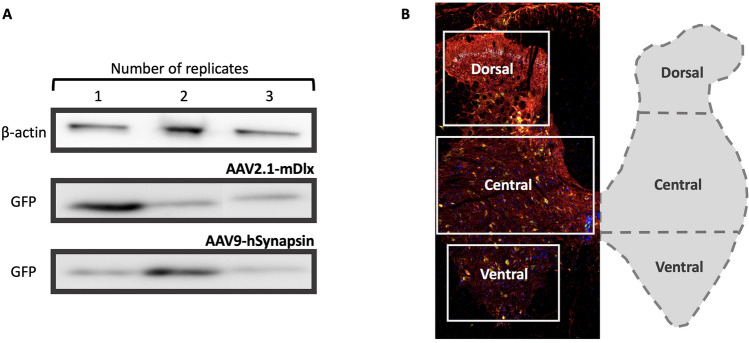
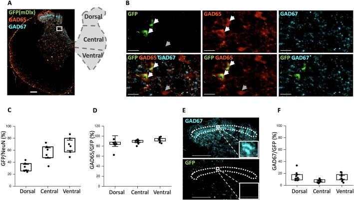
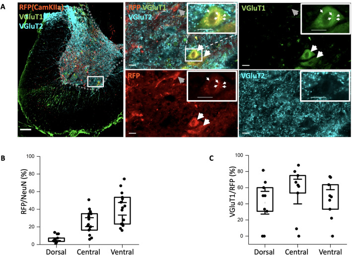
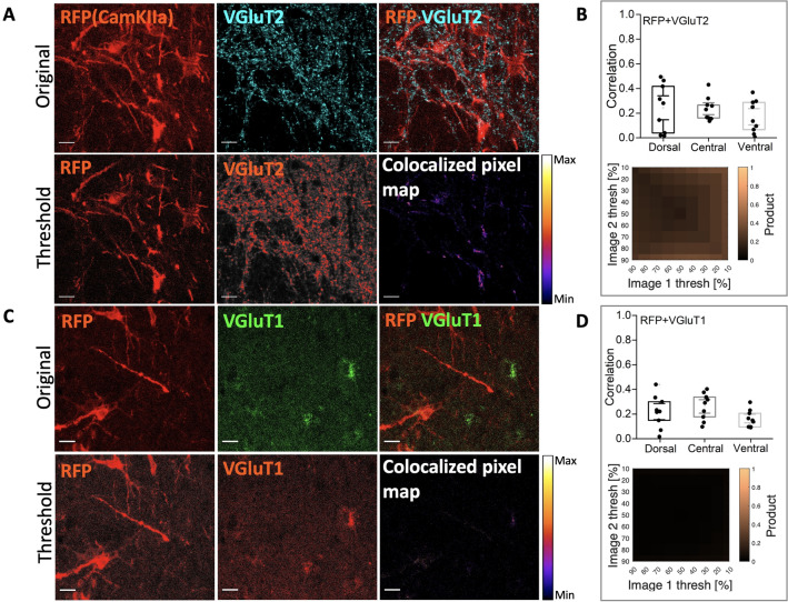
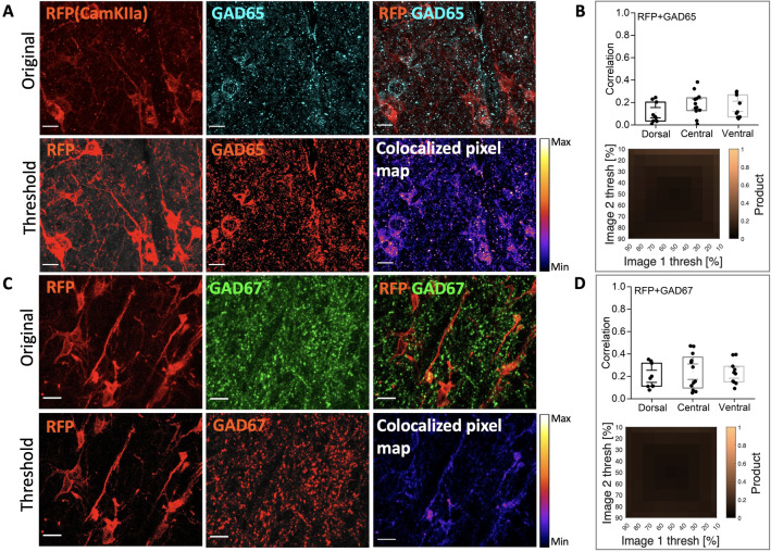
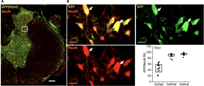
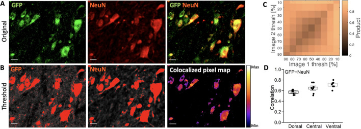
Targeting specific subtypes of interneurons in the spinal cord is primarily restricted to a small group of genetic model animals. Since the development of new transgenic model animals can be expensive and labor intensive, it is often difficult to generalize these findings and verify them in other model organisms, such as the rat, ferret or monkey, that may be more beneficial in certain experimental investigations. Nevertheless, endogenous enhancers and promoters delivered using an adeno-associated virus (AAV) have been successful in providing expression in specific subtypes of neurons in the forebrain of wildtype animals, and therefore may introduce a shortcut. GABAergic interneurons, for instance, have successfully been targeted using the mDlx promoter, which has recently been developed and is now widely used in wild type animals. Here, we test the specificity and efficiency of the mDlx enhancer for robust targeting of inhibitory interneurons in the lumbar spinal cord of wild-type rats using AAV serotype 2 (AAV2). Since this has rarely been done in the spinal cord, we also test the expression and specificity of the CamKIIa and hSynapsin promoters using serotype 9. We found that AAV2-mDlx does in fact target many neurons that contain an enzyme for catalyzing GABA, the GAD-65, with high specificity and a small fraction of neurons containing an isoform, GAD-67. Expression was also seen in some motor neurons although with low correlation. Viral injections using the CamKIIa enhancer via AAV9 infected in some glutamatergic neurons, but also GABAergic neurons, whereas hSynapsin via AAV9 targets almost all the neurons in the lumbar spinal cord.


Conflict of interest statement

The authors declare no competing interests.

Figures

Figure 1
Figure 1
Expression of AAV viruses in the lumbar spinal cord of wild-type Wistar adult rats. (A) Western blots of the images confirming expression of two viruses, AAV2.1-mDlx-GCaMP6f-Fishell-2 (N = 3 rats) and AAV9-hSynapsin-soCoChR-GFP (N = 3 rats) with beta-actin as the reference protein. (B) Confirmation of expression was performed by visual inspection of the fluorescent reporter, where after immunohistochemistry was performed and cell colocalization and counting was performed in three dorsoventral regions of the lumbar cord (D, 560 × 720  μm2), central (C, 590 ×  1070 μm2) and ventral (V, 460 × 770 μm2). Red color represents GAD65, green GFP (mDlx), cyan GAD67 and blue DAPI. Full length western blots are shown in the supplementary Fig.  1A,B.
Figure 2
Figure 2
The AAV2-mDlx enhancer primarily expressed in GAD65-containing neurons in the lumbar spinal cord. (A) Sample tissue section and illustration of the spinal cord showing a distinct layer of GAD67-containing synaptic terminals and neurons (cyan) whereas GAD65-containing neurons are more dispersed (red). The AAV2-mDlx-driven viral expression is shown in green (GFP). (B) A highlighted section (panel A) indicates co-localization of GFP and GAD65, but not GAD67. There are also instances of GAD65+ cells where GFP was not expressed (dim arrow). (C,D) Fraction of co-localization of GFP+ cells and cells containing NeuN (C, N = 2 rats, n = 3 sections/rat) and GAD65 (D, N = 3 animals, n = 3 sections) in the three dorso-ventral regions. (E) The dense layer of GAD67-containing neurons in the dorsal horn (substantia gelatinosa) has little or no overlap with GFP. (F) Cell count (shown as %) of the co-expression in the three regions between GAD67 and GFP positive cells (N = 3 animals, n = 3 sections/animal).
Figure 3
Figure 3
AAV2-mDlx has expression in GABAergic neurons, specifically the GAD65+ neurons. (A) Top row: original images of mDlx reporter (GFP, green) and GAD65 (red) and GAD67 (cyan) and a colocalization pixel map of GFP/GAD65 (right). Middle row: thresholded versions of top row, with the overlap in heat map (right) (scale bar 20 μm). Bottom row: overlap of GFP/GAD65/GAD67, GFP/GAD65 and GFP/GAD67, respectively. (B,C) Varying the threshold and calculating the co-localization as correlation (C). Note N = 4 animals, n = 3 sections/animal.
Figure 4
Figure 4
Infection of spinal neurons with AAV9-CamKIIa primarily in the central and ventral regions. (A) Overview of infection (red, mScarlet designated here as RFP) in the half spinal section combined with the immunostaining of VGluT1 (green) and 2 (cyan) and zoomed in images of a highlighted region in half section and the immunostaining of VGluT1 and 2 (green and cyan), indicates some overlap with VGluT1, whereas the VGluT2 primarily stains the synaptic terminals. (B) Counting neurons (NeuN) that co-expressed with the cells expressing RFP (N = 6 animals, n = 3 sections/animal). (C) Box plot of the co-expressed cell count with VGluT1 positive neurons (N = 3 animals, n = 3 sections/animal). Scale bar 200 μm in image A (left) and 20 μm in the insets.
Figure 5
Figure 5
AAV9-CamKIIa has little or no colocalization with VGluT2 and VGluT1 staining. (A) Top row: the infected cells with the CamKIIa promoter (AAV9) express mScarlet (RFP) and its overlap with VGluT2 (cyan), and second row: thresholded views of the top row and the heat map of RFP and VGluT2. (B) Top right grey box: fluorescent correlation plots of RFP and VGluT2 show very little co-expression. (C) First row: original images showing mScarlet (RFP), VGluT1 (green) and their overlap, and bottom row: the threshold views of the images and and the heat map for co-localization. (D) Bottom right grey box: the correlation for various levels of threshold appear dark. (N = 3 animals, n = 3–4 sections/animal). Scale bar 20 μm.
Figure 6
Figure 6
AAV9-CamKIIa-infected cells has poor overlap with GAD65 and GAD67 biomarkers. (A) Top row: mScarlet expression (red, RFP) and the immunostaining of GAD65 (cyan) has only little co-expression, and second row: threshold representations of the original images and a heat map co-expression plot of RFP/GAD65. (B) Top right grey box: plots show poor correlation in pixel-overlap between RFP and GAD65. (C) First row: original images of RFP, GAD67 (green) and their overlap, and bottom row: threshold representations of the original images and the heat map of RFP/GAD67. (D) Bottom right grey box: the mosaic of correlation coefficients for various thresholds, indicate poor overlap between RFP and GAD67. (N = 3 rats, n = 3-4 sections/rat). Scale bar 20 μm.
Figure 7
Figure 7
AAV9-hSyn1 targets a large fraction of spinal neurons. (A) A spinal section with infection using hSyn1-promoter co-stained with NeuN marker for neurons. (B) Highlighted region in (A) shows the extensive overlap between the infected cells and the NeuN marker and box plots at the bottom right show 40–60% co-expression of NeuN and GFP in the dorsal region whereas 90% overlap in the central and ventral regions. Scale bar = 200 μm in image (A) and 20 μm in the zoomed-in images (B). N = 4 animals, n = 3 sections/animal.
Figure 8
Figure 8
AAV9-hSyn1-driven infection correlation with NeuN biomarker. (A) Top row: expression of GFP with hSyn1 promoter has high overlap with the NeuN staining. (B) Threshold and co-localization in a heat map. (C,D) The co-localization is quantified for different thresholds and across the three regions. Scale bar 20 μm. N = 3 animals, n = 3 sections/animal.

References

    1. Luo L, Callaway EM, Svoboda K. Genetic dissection of neural circuits: A decade of progress. Neuron. 2018;98:256–281. doi: 10.1016/j.neuron.2018.03.040. - DOI - PMC - PubMed
    1. Chettih SN, Harvey CD. Single-neuron perturbations reveal feature-specific competition in V1. Nature. 2019;567:334–340. doi: 10.1038/s41586-019-0997-6. - DOI - PMC - PubMed
    1. Shemesh, O. A. et al. Publisher Correction: Temporally precise single-cell-resolution optogenetics (Nature Neuroscience (2017) 10.1038/s41593-017-0018-8).Nat. Neurosci.21, 896. 10.1038/s41593-018-0097-1 (2018). - PubMed
    1. Kügler S, Kilic E, Bähr M. Human synapsin 1 gene promoter confers highly neuron-specific long-term transgene expression from an adenoviral vector in the adult rat brain depending on the transduced area. Gene Ther. 2003;10:337–347. doi: 10.1038/sj.gt.3301905. - DOI - PubMed
    1. Watakabe A, et al. Comparative analyses of adeno-associated viral vector serotypes 1, 2, 5, 8 and 9 in marmoset, mouse and macaque cerebral cortex. Neurosci. Res. 2015;93:144–157. doi: 10.1016/j.neures.2014.09.002. - DOI - PubMed

Publication types

Supplementary concepts