

Teaching the Virtual Brain
- PMID: 35606668
- PMCID: PMC9712841
- DOI: 10.1007/s10278-022-00652-5
Teaching the Virtual Brain
Abstract
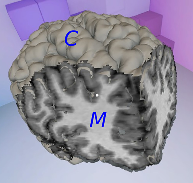
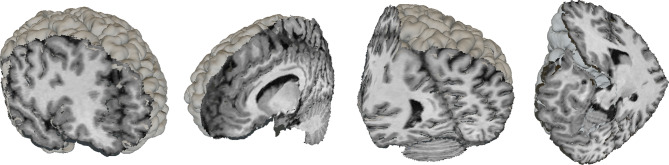
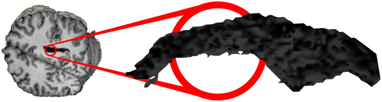
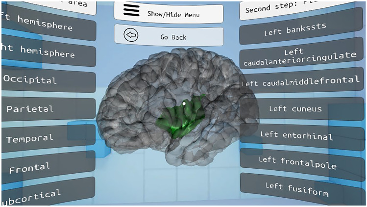
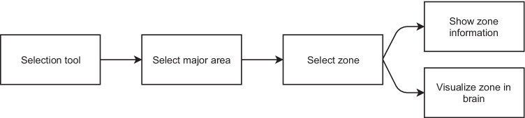
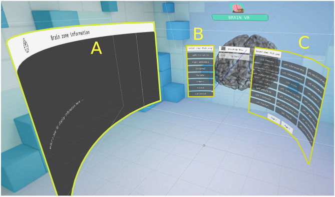
As a complex three-dimensional organ, the inside of a human brain is difficult to properly visualize. Magnetic Resonance Imaging provides an accurate model of the brain of a patient, but its medical or educational analysis as a set of flat slices is not enough to fully grasp its internal structure. A virtual reality application has been developed to generate a complete three-dimensional model based on MRI data, which users can explore internally through random planar cuts and color cluster isolation. An indexed vertex triangulation algorithm has been designed to efficiently display large amounts of complex three-dimensional vertex clusters in simple mobile devices. Feedback from students suggests that the resulting application satisfactorily complements theoretical lectures, as virtual reality allows them to better observe different structures within the human brain.
Keywords: Brain; Brain exploration; Education; Virtual reality.
© 2022. The Author(s).
Conflict of interest statement
The authors declare that they have no conflict of interest.
Figures


References
-
- Huettel SA, Song AW, McCarthy G. Functional Magnetic Resonance Imaging. USA: Freeman; 2009.
-
- Lauterbur P (1973) Image formation by induced local interactions: examples employing nuclear magnetic resonance. Nature 242:190-191 - PubMed
-
- Rinck PA (2019) Magnetic resonance in medicine: a critical introduction. BoD–Books on Demand
-
- Steuer J. Defining virtual reality: Dimensions determining telepresence. Journal of communication. 1992;42(4):73–93. doi: 10.1111/j.1460-2466.1992.tb00812.x. - DOI
-
- Henrysson A, Billinghurst M, Ollila M (2005) Virtual object manipulation using a mobile phone. In: Proceedings of the 2005 international conference on Augmented tele-existence, ACM, pp 164–171
Publication types
MeSH terms
LinkOut - more resources
Full Text Sources
Research Materials
