. 2022 May 23;17(1):210.
doi: 10.1186/s13023-022-02365-y.

Shortcutting the diagnostic odyssey: the multidisciplinary Program for Undiagnosed Rare Diseases in adults (UD-PrOZA)

Collaborators, Affiliations

Shortcutting the diagnostic odyssey: the multidisciplinary Program for Undiagnosed Rare Diseases in adults (UD-PrOZA)

Nika Schuermans et al. Orphanet J Rare Dis. .

Abstract

Background: In order to facilitate the diagnostic process for adult patients suffering from a rare disease, the Undiagnosed Disease Program (UD-PrOZA) was founded in 2015 at the Ghent University Hospital in Belgium. In this study we report the five-year results of our multidisciplinary approach in rare disease diagnostics.

Methods: Patients referred by a healthcare provider, in which an underlying rare disease is likely, qualify for a UD-PrOZA evaluation. UD-PrOZA uses a multidisciplinary clinical approach combined with state-of-the-art genomic technologies in close collaboration with research facilities to diagnose patients.

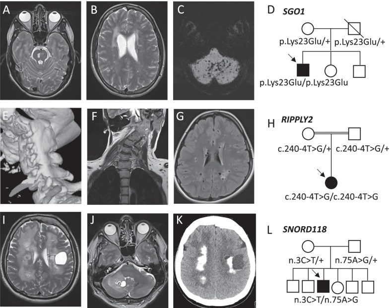
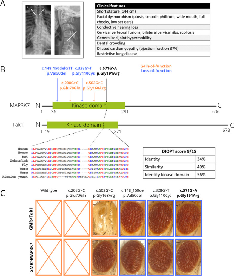
Results: Between 2015 and 2020, 692 patients (94% adults) were referred of which 329 (48%) were accepted for evaluation. In 18% (60 of 329) of the cases a definite diagnosis was made. 88% (53 of 60) of the established diagnoses had a genetic origin. 65% (39 of 60) of the genetic diagnoses were made through whole exome sequencing (WES). The mean time interval between symptom-onset and diagnosis was 19 years. Key observations included novel genotype-phenotype correlations, new variants in known disease genes and the identification of three new disease genes. In 13% (7 of 53), identifying the molecular cause was associated with therapeutic recommendations and in 88% (53 of 60), gene specific genetic counseling was made possible. Actionable secondary findings were reported in 7% (12 of 177) of the patients in which WES was performed.

Conclusion: UD-PrOZA offers an innovative interdisciplinary platform to diagnose rare diseases in adults with previously unexplained medical problems and to facilitate translational research.

Keywords: ACMSD; Diagnostic odyssey; Diagnostic yield; IRF2BPL; MAP3K7; PLAAT3; Rare diseases; SGO1; SNORD118; UD-PrOZA; Whole exome sequencing.


Conflict of interest statement

The authors declare no competing interests.

Figures

Fig. 1
Fig. 1
(Neuro)imaging of patients diagnosed with a rare disease which led to expansion of the phenotypic spectrum or which was associated with therapeutic changes. A, B T2- weighted MRI images of a patient diagnosed with CAID showing pontine and periventricular hyperintensities. C MRI image with maximum intensity projection (MIP) showing diffuse cerebellar microbleeds. D Pedigree of the CAID patient. He is the first child of non-consanguineous parents with a homozygous pathogenic SGO1 variant (p.Lys23Glu). Both parents are heterozygotes. E 3D-reconstruction of CT-scan imaging of the cervical spine revealing a unilateral fusion of the cervical vertebrae C1-C6 in a patient diagnosed with spondylocostal dysostosis 6. F Coronal MRI image showing torticollis. G Axial MRI Flair image showing enlarged perivascular spaces and white matter lesions. H Pedigree of the patient with consanguineous parents and a homozygous RIPPLY2 variant (c.240-4T>G). I, J T2-weighted MRI images of patient 4 revealing the presence of a large cyst in the left cerebral and the right cerebellar hemisphere and diffuse periventricular white matter hyperintensities. K CT-scan axial section showing diffuse cerebral calcifications. L Pedigree of the LCC patient. He is the only affected child of non-consanguineous parents and is compound heterozygous for two variants in SNORD118 (n.3C > T; n.75A > G). Both parents were heterozygous for one pathogenic variant
Fig. 2
Fig. 2
Variant modeling in Drosophila establishes MAP3K7 p.(Gly191Arg) as a novel pathogenic LoF variant causing CSCFS. A Posteroanterior view of a chest X-ray showing scoliosis and bilateral cervical ribs. Lateral view of a cervical spine X-ray shows a fusion of C5 and C6. An overview of all clinical features is provided. B The MAP3K7 and Tak1 protein contain an evolutionary conserved kinase domain. Two of the known GoF variants (associated with FMD2) are depicted in orange, two of the known LoF variants (associated with CSCFS) are represented in blue. Our new variant (c.571G>A, p.Gly191Arg), depicted in black, is located in de kinase domain and replaces a highly conserved glycine for an arginine residue. C Overexpression of wild-type Drosophila Tak1 and human MAP3K7 in the Drosophila eye using a GMR driver resulted in early pupal lethality. Overexpression of fly Tak1 and human MAP3K7 with GoF variant p.Glu70Gln fully recapitulated the phenotype of wild-type overexpression. With the p.Gly168arg variant a discrepancy between the human and fly protein was noted. Overexpression of the fly Tak1 variant resulted in a severe eye phenotype whereas overexpression of the human variant did not visibly affect the eye, pointing towards a milder GoF effect compared to the other GoF mutation. Overexpression of known LoF variants p.Val50del and p.Gly110Cys in human and fly proteins lost the wild-type eye phenotype. Overexpression of our novel variant p.Gly191Arg resulted in a normal eye thereby establishing the LoF nature of this variant

References

    1. Julkowska D, Austin CP, Cutillo CM, Gancberg D, Hager C, Halftermeyer J, et al. The importance of international collaboration for rare diseases research: a European perspective. Gene Ther. 2017;24(9):562–571. doi: 10.1038/gt.2017.29. - DOI - PMC - PubMed
    1. Fernandez-Marmiesse A, Gouveia S, Couce ML. NGS technologies as a turning point in rare disease research. Diagnosis Treat Curr Med Chem. 2018;25(3):404–432. doi: 10.2174/0929867324666170718101946. - DOI - PMC - PubMed
    1. Dawkins HJS, Draghia-Akli R, Lasko P, Lau LPL, Jonker AH, Cutillo CM, et al. Progress in rare diseases research 2010–2016: an IRDiRC perspective. Clin Transl Sci. 2018;11(1):11–20. doi: 10.1111/cts.12501. - DOI - PMC - PubMed
    1. Nguengang Wakap S, Lambert DM, Olry A, Rodwell C, Gueydan C, Lanneau V, et al. Estimating cumulative point prevalence of rare diseases: analysis of the Orphanet database. Eur J Hum Genet. 2020;28(2):165–173. doi: 10.1038/s41431-019-0508-0. - DOI - PMC - PubMed
    1. Rath A, Salamon V, Peixoto S, Hivert V, Laville M, Segrestin B, et al. A systematic literature review of evidence-based clinical practice for rare diseases: what are the perceived and real barriers for improving the evidence and how can they be overcome? Trials. 2017;18(1):556. doi: 10.1186/s13063-017-2287-7. - DOI - PMC - PubMed

Publication types