. 2022 May 23;19(1):114.
doi: 10.1186/s12974-022-02472-4.

A crosstalk between gut and brain in sepsis-induced cognitive decline

Affiliations

A crosstalk between gut and brain in sepsis-induced cognitive decline

Vijayasree V Giridharan et al. J Neuroinflammation. .

Abstract

Background: Sepsis is a potentially fatal disease characterized by acute organ failure that affects more than 30 million people worldwide. Inflammation is strongly associated with sepsis, and patients can experience impairments in memory, concentration, verbal fluency, and executive functioning after being discharged from the hospital. We hypothesize that sepsis disrupts the microbiota-gut-brain axis homeostasis triggering cognitive impairment. This immune activation persists during treatment, causing neurological dysfunction in sepsis survivors.

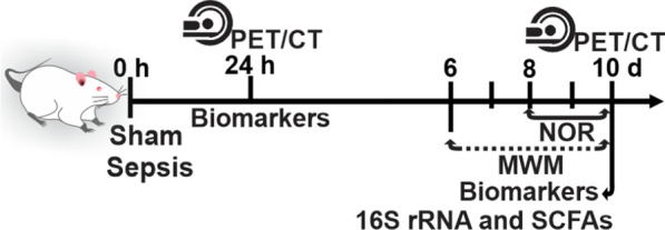
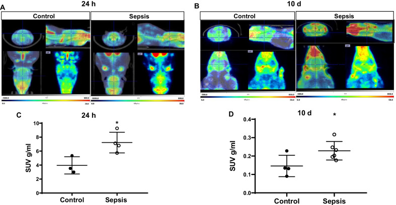
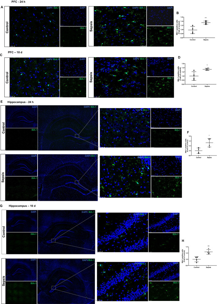
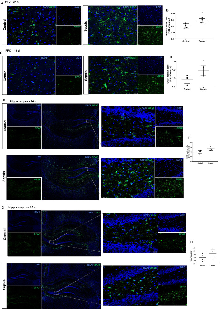
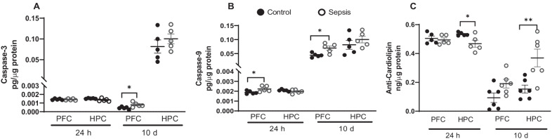
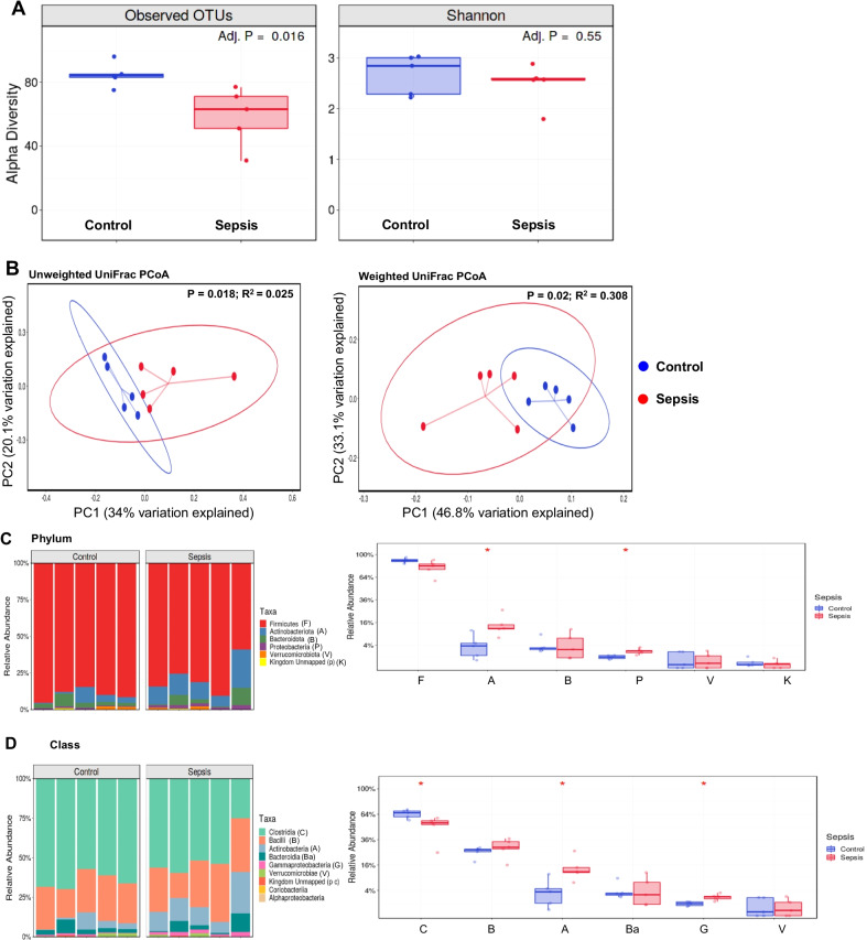
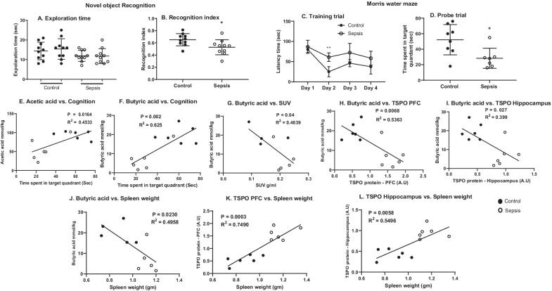
Methods: To test our hypothesis, adult Wistar rats were subjected to cecal-ligation and perforation (CLP) or sham (non-CLP) surgeries. The animals were subjected to the [11C]PBR28 positron emission tomography (PET)/computed tomography (CT) imaging at 24 h and 10 days after CLP and non-CLP surgeries. At 24 h and 10 days after surgery, we evaluated the gut microbiome, bacterial metabolites, cytokines, microglia, and astrocyte markers. Ten days after sepsis induction, the animals were subjected to the novel object recognition (NOR) and the Morris water maze (MWM) test to assess their learning and memory.

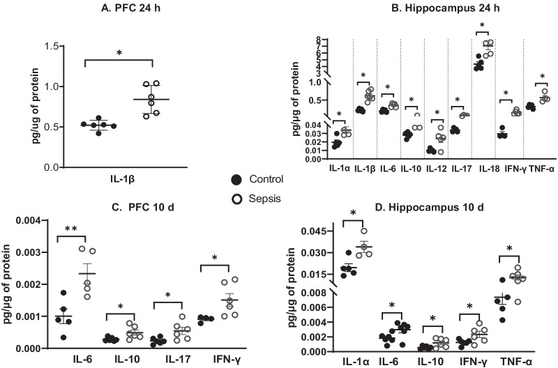
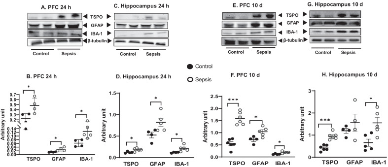
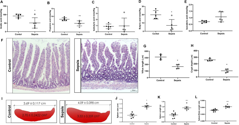
Results: Compared to the control group, the 24-h and 10-day CLP groups showed increased [11C]PBR28 uptake, glial cells count, and cytokine levels in the brain. Results show that sepsis modulates the gut villus length and crypt depth, alpha and beta microbial diversities, and fecal short-chain fatty acids (SCFAs). In addition, sepsis surviving animals showed a significant cognitive decline compared with the control group.

Conclusions: Since several pharmacological studies have failed to prevent cognitive impairment in sepsis survivors, a better understanding of the function of glial cells and gut microbiota can provide new avenues for treating sepsis patients.

Keywords: Astrocyte; Behavior; Cytokines; Inflammation; Microbiome; Microglia; PET; Sepsis; TSPO.


Conflict of interest statement

AP received preclinical research support from ACADIA Pharmaceuticals. All other authors declare that they have no competing interests.

Figures

Fig. 1
Fig. 1
Schematic representation of timeline and experimental design. NOR novel object recognition, MWM morris water maze, 16S rRNA sequencing, and SCFAs short-chain fatty acids
Fig. 2
Fig. 2
In vivo PET imaging using TSPO specific radiotracer [11C]PBR28. Representative summed images from the dynamic reconstruction of simultaneously acquired PET/CT scans from Wistar rats subjected to experimental sepsis by cecal ligation and perforation (CLP) and controls, 60 min after intravenous injection [11C]PBR28. In the experimental sepsis model, the brain’s coronal, sagittal, and dorsal orientations show increased brain [11C]PBR28 uptake than in controls. A Twenty-four hours and B Ten days after experimental sepsis induction. The SUV scale represents the standardized uptake value (SUV). Quantification of [11C]PBR28 uptake detected in the experimental sepsis model was significantly higher compared to control based on measures of brain/muscle. C SUV 24 h after experimental sepsis induction. D SUV 10 days after experimental sepsis induction. The results are expressed as the mean ± SEM for n = 3–6 rats. *p < 0.05 compared to controls
Fig. 3
Fig. 3
BioPlex determined the cytokine levels in the PFC and hippocampus. We evaluated the levels of cytokines (IL1-α, IL1-β, IL-4, IL-6, IL-7, IL-10, IL-12, IL-13, IL-17, IL-18, TNF-α, and INF-γ) in the A PFC and B Hippocampus at 24 h after CLP and non-CLP surgeries. C PFC and D Hippocampus at 10 days after CLP and non-CLP surgeries. The results are expressed as the mean ± SEM for n = 4–6 rats. *p < 0.05 and **p < 0.01 compared to controls
Fig. 4
Fig. 4
Upregulation of glial marker at 24 h A PFC and C Hippocampus; and 10 days E PFC and G Hippocampus after experimental sepsis induction in the Western blot analysis. Quantification of immunoblot data using the densitometric analysis of each protein was carried out using Image Lab™ software (Bio-Rad, California, USA). Representative immunoblots and quantification for 24 h, IBA-1, GFAP, and TSPO in the PFC B and hippocampus D. Representative immunoblots and quantification for 10 days, IBA-1, GFAP, and TSPO in the PFC F, and hippocampus H. The values for all protein levels were normalized to those of β-tubulin. The results are expressed as the mean ± SEM for n = 4–6 rats. *p < 0.05 and ***p < 0.001 compared to controls
Fig. 5
Fig. 5
Increased microglial positive cells at 24 h and 10 days after experimental sepsis induction in PFC and hippocampus. Representative microscopic field images (magnification, × 400) immunostained with IBA-1 antibodies in the PFC A 24 h, B quantification of 24 h, **p < 0.01, C 10 days, D quantification of 10 days, *p < 0.05, after experimental sepsis induction. Representative microscopic field images (magnification, × 100 and × 400) immunostained with IBA-1 antibodies in the hippocampus E 24 h, F Quantification of 24 h, *p < 0.05, G 10 days, H Quantification of 10 days, **p < 0.01, after experimental sepsis induction. The results are expressed as the mean ± SEM for n = 4 rats
Fig. 6
Fig. 6
Astroglial cells at 24 h and 10 days after experimental sepsis induction in PFC and hippocampus. Representative microscopic field images (magnification, × 400) immunostained with GFAP antibodies in the PFC A 24 h, B Quantification of 24 h, *p < 0.05, C 10 days, D quantification of 10 days, *p < 0.05, after experimental sepsis induction. Representative microscopic field images (magnification, × 100 and × 400) immunostained with GFAP antibodies in the hippocampus E 24 h, F Quantification of 24 h, *p < 0.05, G 10 days, H Quantification of 10 days, p ≥ 0.05, after experimental sepsis induction. The results are expressed as the mean ± SEM for n = 4 rats
Fig. 7
Fig. 7
Cardiolipin and caspase levels at 24 h and 10 days after experimental sepsis were measured using ELISA in PFC and hippocampus. At 24 h and 10 days, tissue protein levels of A caspase-3, B caspase-9, and C. cardiolipin at PFC and hippocampus in experimental sepsis and sham control rats. The results are expressed as the mean ± SEM for n = 5–6 rats. *p < 0.05 and **p < 0.01 compared to controls
Fig. 8
Fig. 8
Results from 16S rRNA sequencing. A Alpha diversity, Observed OTUs adjusted p = 0.016 and Shannon index adjusted p = 0.55. B Beta diversity, unweighted unifrac PCoA p = 0.018; weighted unifrac PCoA p = 0.02. C Relative abundance in phyla. The phylum Actinobacteria *p < 0.05, and Proteobacteria *p < 0.05 increased in the sepsis group. D Relative abundance in class. The class Clostridia decreased *p < 0.05, Actinobacteria *p < 0.05 and Gammaproteobacteria *p < 0.05 classes increased in the sepsis group. The results are expressed as the mean ± SEM for n = 5 rats
Fig. 9
Fig. 9
SCFAs evaluation. The levels of SCFAs were measured from cecum fecal samples A Acetic acid, B Propionic acid, C Isobutyric acid, D Butyric acid, and E Isovaleric acid. The levels of acetic acid, *p < 0.05; propionic acid, **p < 0.01; and butyric acid, *p < 0.05 significantly reduced after sepsis. The results are expressed as the mean ± SEM for n = 5 rats. F Gut H&E staining G Villus length, *p < 0.05, and H Crypt depth, *p < 0.05 decreased at 10 days after the sepsis (scale bar = 100 μm; magnification, × 100) The results are expressed as the mean ± SEM for n = 4 rats. I and J Spleen size, K Spleen weight, and L Spleen/body weight ratio, ***p < 0.001, significantly increased 10 days after sepsis. The results are expressed as the mean ± SEM for n = 7–8 rats
Fig. 10
Fig. 10
Ten days after the induction of experimental sepsis, Wistar rats were subjected to novel object recognition (NOR) test. A Locomotor activity and B. Recognition index. The recognition index decreased in the sepsis group compared to the control group, *p < 0.05. Evaluation of spatial memory task by Morris water maze. C Training trial, and D Probe trial. On day 2 of the training trial sepsis group taken more time to reach the platform as compared to control group, **p < 0.01. In the probe trial, sepsis-subjected rats demonstrated less time spent in the target quadrant, *p < 0.05, and n = 7–10 rats. The Pearson correlation between E Acetic acid vs. cognition, F Butyric acid vs. cognition, G Butyric acid vs. SUV uptake, H Butyric acid vs. TSPO at PFC, and I Butyric acid vs. TSPO in hippocampus, J Spleen weight vs. butyric acid, K Spleen weight vs. TSPO at PFC, L Spleen weight vs. TSPO at hippocampus, n = 4–6 rats. The results are expressed as the mean ± SEM

References

    1. CDC, Data & Reports-Sepsis, C.f.D.C.a. Prevention., Editor. updated 2018.
    1. Prescott HC, Angus DC. Enhancing recovery from sepsis: a review. JAMA. 2018;319(1):62–75. doi: 10.1001/jama.2017.17687. - DOI - PMC - PubMed
    1. Singer M, et al. The third international consensus definitions for sepsis and septic shock (sepsis-3) JAMA. 2016;315(8):801–810. doi: 10.1001/jama.2016.0287. - DOI - PMC - PubMed
    1. Iwashyna TJ, et al. Long-term cognitive impairment and functional disability among survivors of severe sepsis. JAMA J Am Med Assoc. 2010;304(16):1787–1794. doi: 10.1001/jama.2010.1553. - DOI - PMC - PubMed
    1. Iwashyna TJ, et al. Population burden of long-term survivorship after severe sepsis in Older Americans. J Am Geriatr Soc. 2012;60(6):1070–1077. doi: 10.1111/j.1532-5415.2012.03989.x. - DOI - PMC - PubMed