

Structural comparisons reveal diverse binding modes between nucleosome assembly proteins and histones
- PMID: 35606827
- PMCID: PMC9128123
- DOI: 10.1186/s13072-022-00452-9
Structural comparisons reveal diverse binding modes between nucleosome assembly proteins and histones
Abstract
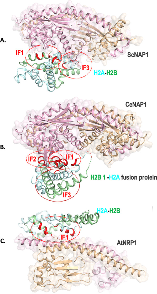
Nucleosome assembly proteins (NAPs) are histone chaperones that play a central role in facilitating chromatin assembly/disassembly which is of fundamental importance for DNA replication, gene expression regulation, and progression through the cell cycle. In vitro, NAPs bind to the core histones H2A, H2B, H3, H4 and possibly to H1. The NAP family contains well-characterized and dedicated histone chaperone domain called the NAP domain, and the NAP-histone interactions are key to deciphering chromatin assembly. Our comparative structural analysis of the three three-dimensional structures of NAPs from S. cerevisiae, C. elegans, and A. thaliana in complex with the histone H2A-H2B dimer reveals distinct and diverse binding of NAPs with histones. The three NAPs employ distinct surfaces for recognizing the H2A-H2B dimer and vice versa. Though histones are highly conserved across species they display diverse footprints on NAPs. Our analysis indicates that understanding of NAPs and their interaction with histone H2A-H2B remains sparse. Due to divergent knowledge from the current structures analyzed here, investigations into the dynamic nature of NAP-histone interactions are warranted.
Keywords: Histone chaperone; NAP; Nucleosome assembly protein; Structural analysis.
© 2022. The Author(s).
Conflict of interest statement
The authors declare that they have no competing interests.
Figures
Similar articles
-
Structural Insights into the Association of Hif1 with Histones H2A-H2B Dimer and H3-H4 Tetramer.Structure. 2016 Oct 4;24(10):1810-1820. doi: 10.1016/j.str.2016.08.001. Epub 2016 Sep 8. Structure. 2016. PMID: 27618665
-
Distinct roles for histone chaperones in the deposition of Htz1 in chromatin.Biosci Rep. 2014 Sep 19;34(5):e00139. doi: 10.1042/BSR20140092. Biosci Rep. 2014. PMID: 25338502 Free PMC article.
-
Mechanistic and structural insights into histone H2A-H2B chaperone in chromatin regulation.Biochem J. 2020 Sep 18;477(17):3367-3386. doi: 10.1042/BCJ20190852. Biochem J. 2020. PMID: 32941645 Review.
-
Structure, localization and histone binding properties of nuclear-associated nucleosome assembly protein from Plasmodium falciparum.Malar J. 2010 Apr 8;9:90. doi: 10.1186/1475-2875-9-90. Malar J. 2010. PMID: 20377878 Free PMC article.
-
All roads lead to chromatin: multiple pathways for histone deposition.Biochim Biophys Acta. 2013 Mar-Apr;1819(3-4):238-46. Biochim Biophys Acta. 2013. PMID: 24459726 Review.
Cited by
-
Testis- specific Y-encoded- like protein 1 and cholesterol metabolism: Regulation of CYP1B1 expression through Wnt signaling.Front Pharmacol. 2022 Nov 28;13:1047318. doi: 10.3389/fphar.2022.1047318. eCollection 2022. Front Pharmacol. 2022. PMID: 36518674 Free PMC article.
-
Chaperoning the chaperones: Proteomic analysis of the SMN complex reveals conserved and etiologic connections to the proteostasis network.bioRxiv [Preprint]. 2024 May 16:2024.05.15.594402. doi: 10.1101/2024.05.15.594402. bioRxiv. 2024. Update in: Front RNA Res. 2024;2:1448194. doi: 10.3389/frnar.2024.1448194. PMID: 38903116 Free PMC article. Updated. Preprint.
-
Overlap in oncogenic and pro-inflammatory pathways associated with areca nut and nicotine exposure.Cancer Pathog Ther. 2023 Sep 17;2(3):187-194. doi: 10.1016/j.cpt.2023.09.003. eCollection 2024 Jul. Cancer Pathog Ther. 2023. PMID: 39027148 Free PMC article.
-
Single-molecule study reveals Hmo1, not Hho1, promotes chromatin assembly in budding yeast.mBio. 2023 Aug 31;14(4):e0099323. doi: 10.1128/mbio.00993-23. Epub 2023 Jul 11. mBio. 2023. PMID: 37432033 Free PMC article.
-
The Histone Chaperone Network Is Highly Conserved in Physarum polycephalum.Int J Mol Sci. 2023 Jan 5;24(2):1051. doi: 10.3390/ijms24021051. Int J Mol Sci. 2023. PMID: 36674565 Free PMC article.
References
-
- Luger K. Nucleosomes: structure and function. In: Wiley J, editor. eLS. Hoboken: Wiley; 2001.
Publication types
MeSH terms
Substances
LinkOut - more resources
Full Text Sources
Molecular Biology Databases
Research Materials
