. 2022 May 23;20(1):238.
doi: 10.1186/s12967-022-03441-4.

Serum proteome alterations during conventional and extracorporeal resuscitation in pigs

Affiliations

Serum proteome alterations during conventional and extracorporeal resuscitation in pigs

Patrick Bernhard et al. J Transl Med. .

Abstract

Background: Only a small number of patients survive an out-of-hospital cardiac arrest (CA) and can be discharged from hospital alive with a large percentage of these patients retaining neurological impairments. In recent years, extracorporeal cardiopulmonary resuscitation (ECPR) has emerged as a beneficial strategy to optimize cardiac arrest treatment. However, ECPR is still associated with various complications. To reduce these problems, a profound understanding of the underlying mechanisms is required. This study aims to investigate the effects of CA, conventional cardiopulmonary resuscitation (CPR) and ECPR using a whole-body reperfusion protocol (controlled and automated reperfusion of the whole body-CARL) on the serum proteome profiles in a pig model of refractory CA.

Methods: N = 7 pigs underwent 5 min of untreated CA followed by 30 min CPR and 120 min perfusion with CARL. Blood samples for proteomic analysis were drawn at baseline, after CPR and at the end of the CARL period. Following albumin-depletion, proteomic analysis was performed using liquid chromatography-tandem mass spectrometry.

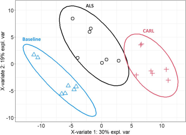
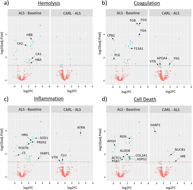
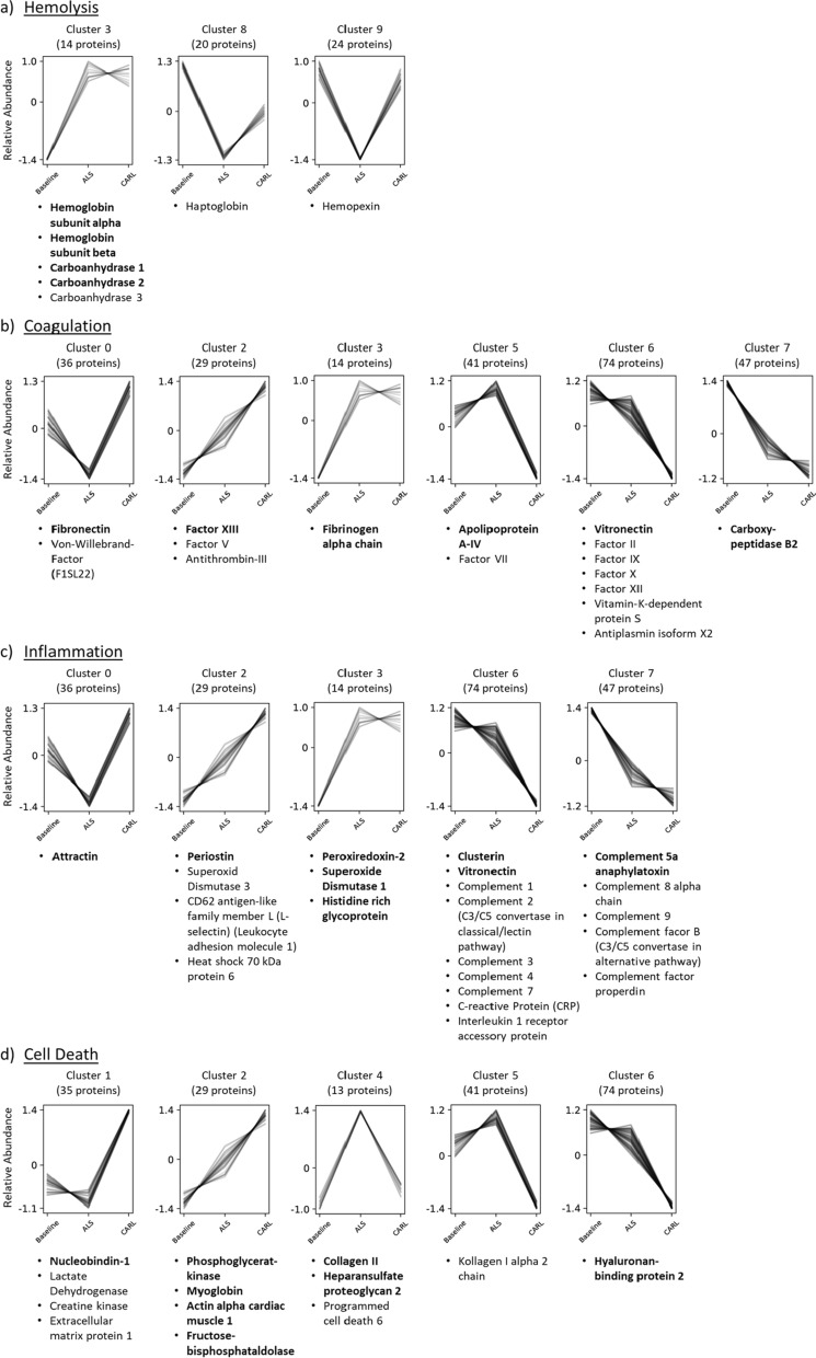
Results: N = 21 serum samples were measured resulting in the identification and quantification of 308-360 proteins per sample and 388 unique proteins in total. The three serum proteome profiles at the investigated time points clustered individually and segregated almost completely when considering a 90% confidence interval. Differential expression analysis showed significant abundance changes in 27 proteins between baseline and after CPR and in 9 proteins after CARL compared to CPR. Significant findings were further validated through a co-abundance cluster analysis corroborating the observed abundance changes.

Conclusions: The presented data highlight the impact of systemic ischemia and reperfusion on the entire serum proteome during resuscitation with a special focus on changes regarding haemolysis, coagulation, inflammation, and cell-death processes. Generally, the observed changes contribute to post-ischemic complications. Better understanding of the underlying mechanisms during CA and resuscitation may help to limit these complications and improve therapeutic options.

Keywords: Cardiopulmonary resuscitation; Extracorporeal cardiopulmonary resuscitation; Extracorporeal membrane oxygenation; Ischemia reperfusion injury; Proteome.


Conflict of interest statement

FB, CB and GT are shareholders in Resuscitec GmbH, Freiburg, Germany, which is a start-up company from the University Medical Centre Freiburg. JSP, SJB and CS are part-time employees of Resuscitec GmbH, Freiburg, Germany. The remaining authors have disclosed that they do not have any conflicts of interest.

Figures

Fig. 1
Fig. 1
Schematic experimental design and proteomics workflow. Pigs underwent a successive series of treatments including cardiac arrest (CA), cardiopulmonary resuscitation (CPR), basic life support (BLS), advanced life support (ALS) and extracorporeal cardiopulmonary resuscitation (ECPR) using CARL (controlled automated reperfusion of the whole body). Blood sampling was performed at the three marked time points including “Baseline” (time point 1), “after ALS” (time point 2) and “after CARL” (time point 3). Subsequent proteomic sample processing involved depletion of albumin and immunoglobulin G (IgG), proteolytic digest and peptide clean-up before samples were subjected to liquid chromatography–tandem mass spectrometry (LC–MS/MS) measurement. VF, ventricular fibrillation
Fig. 2
Fig. 2
Partial least squares discriminating analysis (PLSDA). Protein profiles of each sample, together with the corresponding time point annotation (Baseline, ALS, CARL) were submitted to PLSDA analysis. Ellipses represent an 90% confidence interval
Fig. 3
Fig. 3
Differential expression analysis using pairwise multigroup limma. The volcano plots ad show the results of the same pairwise differential expression analysis between “ALS-Baseline” and “CARL-ALS”. The log2 fold changes (log2FC) are plotted on the x-axis and corresponding adjusted p-values in − log10 scale is shown on the y-axis. The applied adjusted p-value cut-off was set to 0.05 (1.3 in – log10 scale), which is depicted as dashed horizontal line. Each plot highlights significantly up- or downregulated proteins (blue dots) of the respective biological process: a Hemolysis, b Coagulation, c Inflammation, d Cell Death. Hereby, a log2FC > 0 corresponds to an upregulation in the first-mentioned condition. Protein datapoints are labelled with corresponding gene names (black arrows) retrieved from UniProt database (Additional file 4: Table S2)
Fig. 4
Fig. 4
Protein hits and their assigned co-abundance cluster for each considered biological process. Depending on the measured intensities at different time points of the experiment (Baseline, ALS, CARL), the identified proteins were assigned to different co-abundance cluster with a confidence interval of 95%. Cluster assignment was performed by using the Clust algorithm and assigned proteins were manually annotated to one of the respective biological processes: a hemolysis, b coagulation, c inflammation, d cell death. Each line represents an individual protein, while y-axis illustrates the relative abundance change after Z-score normalization. Bold protein names represent proteins that already showed a significant abundance change (adjusted p-value ≤ 0.05) during to the previous differential expression analysis (Fig. 3) for at least one of the considered comparisons. The number of assigned proteins per cluster is shown above each graph

References

    1. Gräsner J-T, Lefering R, Koster RW, Masterson S, Böttiger BW, Herlitz J, et al. EuReCa ONE-27 Nations, ONE Europe. ONE Registry Resusc. 2016;105:188–195. doi: 10.1016/j.resuscitation.2016.06.004. - DOI - PubMed
    1. Gräsner J-T, Wnent J, Herlitz J, Perkins GD, Lefering R, Tjelmeland I, et al. Survival after out-of-hospital cardiac arrest in Europe—results of the EuReCa TWO study. Resuscitation. 2020;148:218–226. doi: 10.1016/j.resuscitation.2019.12.042. - DOI - PubMed
    1. Lascarrou J-B, Merdji H, Le Gouge A, Colin G, Grillet G, Girardie P, et al. Targeted temperature management for cardiac arrest with nonshockable rhythm. N Engl J Med. 2019;381:2327–2337. doi: 10.1056/NEJMoa1906661. - DOI - PubMed
    1. Guy A, Kawano T, Besserer F, Scheuermeyer F, Kanji HD, Christenson J, et al. The relationship between no-flow interval and survival with favourable neurological outcome in out-of-hospital cardiac arrest: implications for outcomes and ECPR eligibility. Resuscitation. 2020;155:219–225. doi: 10.1016/j.resuscitation.2020.06.009. - DOI - PubMed
    1. Perkins GD, Neumar R, Monsieurs KG, Lim SH, Castren M, Nolan JP, et al. The International Liaison Committee on Resuscitation—review of the last 25 years and vision for the future. Resuscitation. 2017;121:104–116. doi: 10.1016/j.resuscitation.2017.09.029. - DOI - PubMed

Publication types