. 2022 May 14:2022:5783867.
doi: 10.1155/2022/5783867. eCollection 2022.

Duchesnea indica Extract Ameliorates LPS-Induced Septic Shock in Mice

Affiliations

Duchesnea indica Extract Ameliorates LPS-Induced Septic Shock in Mice

Yuan Yee Lee et al. Evid Based Complement Alternat Med. .

Abstract

Objective: Duchesnea indica has been reported for its anti-inflammatory properties. However, its efficacy in sepsis has yet to be reported. In this study, we studied the ability of Duchesnea indica extract (DIE) to rescue mice from septic shock and sepsis.

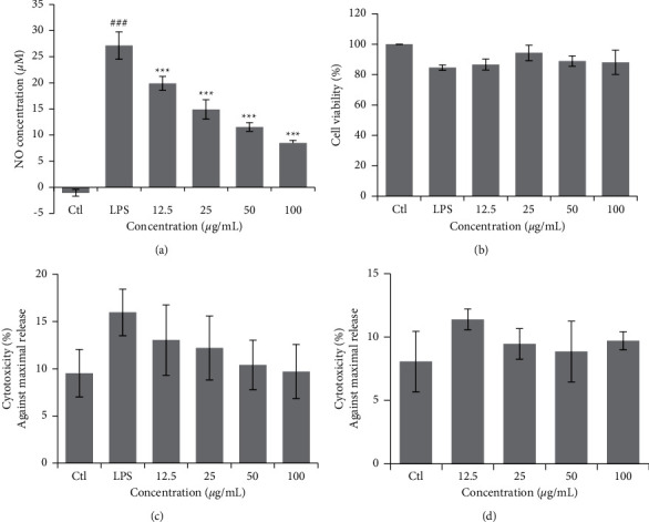
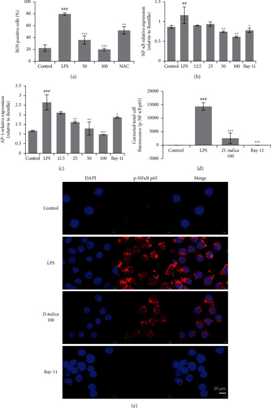
Methods: In vitro studies included the measurement of secreted nitric oxide, cell viability, gene and protein expression via real-time polymerase chain reaction and western blot, and confocal microscopy in RAW 264.7 cells. In vivo studies include a model of septic shock and sepsis in BALB/c mice induced by a lethal and sub-lethal dose of lipopolysaccharide (LPS).

Results: DIE suppressed the expression of proinflammatory cytokines induced by LPS and prevented the translocation of NFκB into the nucleus of RAW 264.7 cells. It also prevented reactive oxygen species damage induced by LPS in murine bone marrow-derived macrophages. Models of sepsis and septic shock were established in BALB/c mice and DIE-rescued mice from septic shock. DIE also reversed the increase in tumor necrosis factor-α and nitrite levels in the serum of mice induced with sepsis. DIE also prevented the translocation of NFκB from the cytosol into the nucleus in murine lungs. Histopathological damage induced by sepsis was reversed in the testis, liver, and lungs of mice.

Conclusion: In conclusion, DIE is a suitable candidate for development as a therapeutic agent for sepsis.


Conflict of interest statement

All of the authors declare no competing interests.

Figures

Figure 1
Figure 1
UPLC-QT of MS analysis of DIE. DIE was analyzed by UPLC-QToF-MS to identify the components. Peaks were compared to a standard of ellagic acid.
Figure 2
Figure 2
DIE suppresses LPS-induced NO production without cytotoxicity in RAW 264.7 cells. RAW 264.7 cells were seeded into 24-well plates and incubated for 24 h before treatment with DIE and LPS. Supernatants were collected after 18 h for NO analysis using the Griess reagent (a). MTT was added to the cells for 3 h and the crystals were dissolved with DMSO (b). Lactate dehydrogenase (LDH) activity was detected in RAW 264.7 cells with an LDH assay kit with LPS (c) or without LPS (d). Experiments were repeated in triplicates. Data were presented as the mean ± SD. # was p < 0.05, ## indicates p < 0.01, and ### indicates p < 0.001 as compared with the control group, whereas was p < 0.05, ∗∗ indicates p < 0.01, and ∗∗∗ indicates p < 0.001 as compared with the LPS-treated group.
Figure 3
Figure 3
Proinflammatory mediators and cytokines in RAW 264.7 cells were suppressed by DIE. RNA was isolated from RAW 264.7 cells using TRIzoL solution. RNA was separated by choloform extraction followed by purification with alcohol. The concentration of RNA was quantified using nanophotometer and reverse transcription was carried out to synthesize cDNA. PCR was done using the cDNA with gene-specific primers. The PCR products were run on ethidium bromide-stained agarose gels and viewed with a gel developer (a). PCR was carried out in triplicate and gels were quantified using ImageJ software (b, c). Relative real-time PCR expression was normalized to the housekeeping gene, GAPDH (d–h). Full gel images were included in the additional information. Experiments were repeated in triplicates. Data are presented as mean ± SD. # was p < 0.05, ## indicates p < 0.01, and ### indicates p < 0.001 as compared with the control group, whereas was p < 0.05, ∗∗ indicates p < 0.01, and ∗∗∗ indicates p < 0.001 as compared with the LPS-treated group.
Figure 4
Figure 4
DIE suppresses protein expression in the NFκB and MAPK pathways in RAW 264.7 cells. Proteins that were extracted from RAW 264.7 cells were separated using 10% SDS–PAGE followed by a transfer onto PVDF membranes. Membranes were blocked with skim milk and incubated with corresponding primary antibodies overnight. Membranes were then incubated with secondary antibody and developed. Representative gel images for the NFκB and MAPK pathways are shown (a, c). Western blot analysis was done in triplicate and quantified using ImageJ software. Proteins in the NFκB pathway were quantified and normalized against the housekeeping gene, β-actin (b), whereas proteins in the MAPK pathway were normalized against their total forms (d). Full blot images were included in the additional information. Experiments were repeated in triplicate. Data are presented as mean ± SD. # was p < 0.05, ## indicates p < 0.01, and ### indicates p < 0.001 as compared with the control group, whereas was p < 0.05, ∗∗ indicates p < 0.01, and ∗∗∗ indicates p < 0.001 as compared with the LPS-treated group.
Figure 5
Figure 5
DIE attenuates oxidative damage and prevents the translocation of phospho-NFκB in RAW 264.7 cells. Bone marrow cells were isolated and differentiated into BMDM. LPS was administered to induce oxidative stress and ROS was measured using DCFDA. Flow cytometry was carried out for quantification (a). pNFκB-Luc and pAP-1-Luc were transfected into RAW 264.7 cells. Cells that were treated with LPS were analyzed for the expression of phospho-NFκB and AP-1 by luciferase assay (b, c). Confocal microscopy was used to investigate the translocation of phospho-NFκB p65 in RAW 264.7 cells. Fluorescence quantification was carried out with ImageJ software and the corrected total cell fluorescence was calculated (d). Images of confocal microscopy where the scale bar represents 10 μm (e). Data are presented as mean ± SD. # was p < 0.05, ## indicates p < 0.01, and ### indicates p < 0.001 as compared with the control group, whereas was p < 0.05, ∗∗ indicates p < 0.01, and ∗∗∗ indicates p < 0.001 as compared with the model group.
Figure 6
Figure 6
DIE rescues mice induced with septic shock and attenuates inflammatory markers in mice. Mice treated with DIE and dexamethasone (Dexa) were administered a single dose of LPS i.p. after 7 days of oral treatment. The mortality of the mice was monitored (a) (n = 10 per group). Serum TNF-α levels and nitrite concentration were determined using an ELISA assay (b, c). Lung tissues were harvested after euthanization. The expression of phospho-NFκB in the nucleus and total NFκB in the cytosol of lung tissue were investigated by western blot analysis. The cytosolic fraction was detected for PARP to ensure purity of the separated fractions (d). Blots were quantified using ImageJ software and the expression in the nucleus was normalized against PARP and the expression in the cytosol was normalized against β-Actin (e) and (f). Full blot images were included in the additional information. Data are presented as mean ± SD. # indicates p < 0.05, ## indicates p < 0.01, and ### indicates p < 0.001 as compared with the control group, whereas was p < 0.05, ∗∗ indicates p < 0.01, and ∗∗∗ indicates p < 0.001 as compared with the LPS-treated group.
Figure 7
Figure 7
DIE prevents histopathological damage in mice induced in a chronic model of sepsis. After euthanization, the testis (a), liver (b), and lungs (c) were directly fixed in 10% neutral buffered formalin and stained with H&E. The respective magnification of the testis was 200x and 100x for the lungs and the liver. The scale bar for the testis group indicates 100 μm, whereas the scale bars for the liver and lungs indicate 200 μm. The arrow indicates the destruction of the seminiferous tubular margin and diminished sperm flagella in the testis. Dexamethasone was indicated as Dexa.

Similar articles

Cited by

References

    1. Singer M., Deutschman C. S., Seymour C. W., et al. The third international consensus definitions for sepsis and septic shock (Sepsis-3) JAMA . 2016;315(8):801–810. doi: 10.1001/jama.2016.0287. - DOI - PMC - PubMed
    1. Lumley J. W., Siu S. K., Rllay S. P., et al. Single dose ceftriaxone as prophylaxis for sepsis in colorectal surgery. ANZ Journal of Surgery . 1992;62(4):292–296. doi: 10.1111/j.1445-2197.1992.tb07559.x. - DOI - PubMed
    1. Eykyn S. J., Phillips I. Metronidazole and anaerobic sepsis. British Medical Journal . 1976;2(6049):1418–1421. doi: 10.1136/bmj.2.6049.1418. - DOI - PMC - PubMed
    1. Zhao Z., Wang B., Mu L., et al. Long-term exposure to ceftriaxone sodium induces alteration of gut microbiota accompanied by abnormal behaviors in mice. Frontiers in Cellular and Infection Microbiology . 2020;10:p. 258. doi: 10.3389/fcimb.2020.00258. - DOI - PMC - PubMed
    1. Wang L., Li S., Liu H., Bao L. Advances in research on the effects of natural drugs with immune-promoting effects on immune function. European Journal of Inflammation . 2020;18 doi: 10.1177/2058739220926878.205873922092687 - DOI

LinkOut - more resources