

Multiple Primary Malignancies With Hypercalcemia Presentation: A Case Report
- PMID: 35607588
- PMCID: PMC9123354
- DOI: 10.7759/cureus.24266
Multiple Primary Malignancies With Hypercalcemia Presentation: A Case Report
Abstract
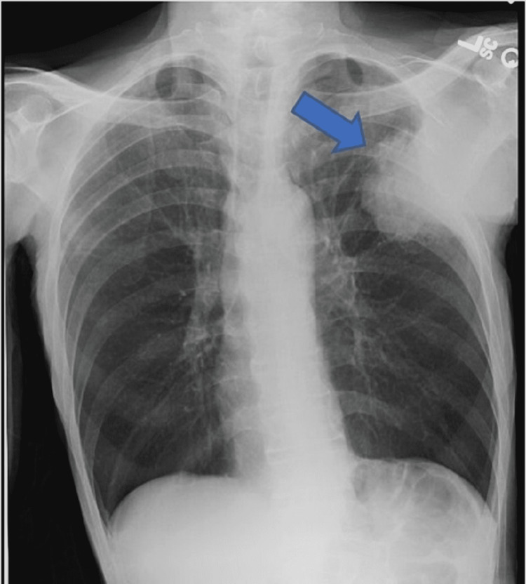
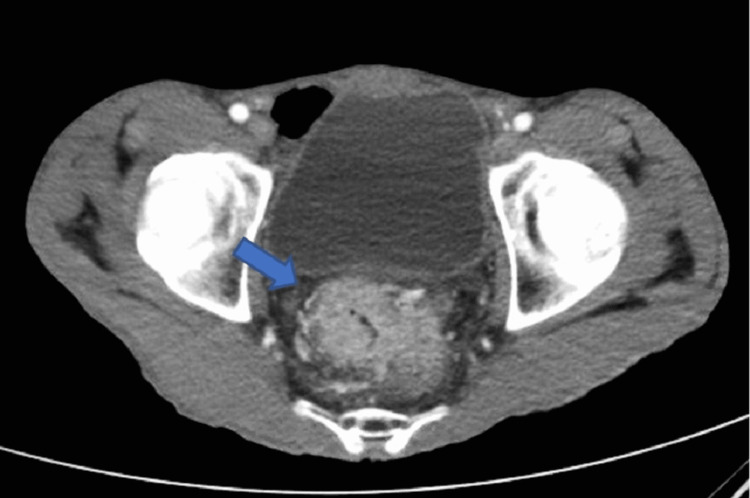
Multiple primary malignancies (MPMs) are defined as having more than one primary malignancy and when each tumor is histologically distinct and unrelated to the others. Multiple risk factors have been found to be associated with MPMs. These include familial syndromes, sequela from treatments of previous malignancies, and environmental factors such as smoking, alcohol consumption, obesity, and male sex. Hypercalcemia has a well-known association with malignancy and is often the first abnormality that leads to further testing. Lung cancer followed by colorectal cancer has the highest mortality of all cancers in the USA, with adenocarcinoma being the most prevalent histological subtype. Further, estimates show that those with one malignancy have a 1.29 times higher risk of developing another malignancy. Hereby, we present a case of a patient with squamous cell carcinoma of the lung who presented with hypercalcemia and incidentally was found to have another primary adenocarcinoma of the colon.
Keywords: colorectal cancer; lung squamous cell carcinoma; multiple primary cancers; paraneoplastic hypercalcemia; rectal adenocarcinoma.
Copyright © 2022, Diraviam et al.
Conflict of interest statement
The authors have declared that no competing interests exist.
Figures


Similar articles
-
Humoral hypercalcemia complicating adenocarcinoma of the sigmoid colon: report of a case.Surg Today. 2005;35(8):692-5. doi: 10.1007/s00595-004-2974-3. Surg Today. 2005. PMID: 16034553 Review.
-
Adenosquamous carcinoma of the colon presenting with hypercalcemia.Cancer. 1987 Sep 1;60(5):1111-6. doi: 10.1002/1097-0142(19870901)60:5<1111::aid-cncr2820600532>3.0.co;2-1. Cancer. 1987. PMID: 3300949 Review.
-
Hypercalcemia of Malignancy: An Atypical Presentation of Endometrial Carcinoma.Cureus. 2024 Sep 24;16(9):e70115. doi: 10.7759/cureus.70115. eCollection 2024 Sep. Cureus. 2024. PMID: 39449942 Free PMC article.
-
Paraneoplastic hypercalcemia.Top Companion Anim Med. 2012 Nov;27(4):156-8. doi: 10.1053/j.tcam.2012.09.003. Top Companion Anim Med. 2012. PMID: 23415382 Review.
-
Paraneoplastic hypercalcemia in a patient with adenosquamous cancer of the colon.Am Surg. 2001 Jun;67(6):585-8. Am Surg. 2001. PMID: 11409809 Review.
References
-
- Multiple primary neoplasms at a single institution: differences between synchronous and metachronous neoplasms. Aydiner A, Karadeniz A, Uygun K, Tas S, Tas F, Disci R, Topuz E. Am J Clin Oncol. 2000;23:364–370. - PubMed
-
- Multiple primary tumors over a lifetime. Copur MS, Manapuram S. http://www.ncbi.nlm.nih.gov/pubmed/31365752. Oncology (Williston Park) 2019;33 - PubMed
Publication types
LinkOut - more resources
Full Text Sources