

doi: 10.1016/j.acpath.2022.100028.
eCollection 2022.
Educational Case: Cranial hemorrhage and traumatic brain injury
Affiliations
- PMID: 35607600
- PMCID: PMC9123211
- DOI: 10.1016/j.acpath.2022.100028
Item in Clipboard
Educational Case: Cranial hemorrhage and traumatic brain injury
Acad Pathol.
.
No abstract available
Keywords: Central nervous system; Cranial hemorrhage; Epidural; Ischemia of the brain; Organ system pathology; Pathology competencies; Subdural; Traumatic brain injury.
Conflict of interest statement
The author(s) declared no potential conflicts of interest with respect to the research, authorship, and/or publication of this article.
Figures

Axial noncontrast CT scan of the brain, right-sided epidural hemorrhage. Source: Reproduced from Radiopaedia.org , rID: 4458, case courtesy of Dr. Sandeep Bhuta, under the Creative Commons Attribution BY-NC-SA 3.0.

Axial noncontrast CT scan of the brain, left-sided subdural hematoma. Source: Reproduced from Radiopaedia.org , rID:71635, courtesy of Dr. Adam Eid Ramsey, under the Creative Commons Attribution BY-NC-SA 3.0.

Representation of the locations of the main types of cerebral hemorrhage.

Axial noncontrast head CT scan, traumatic subarachnoid hemorrhage within the right Sylvian fissure with a contusion. Source: Reproduced from Radiopaedia.org , rID:4852, case courtesy of Assoc Prof Frank Gaillard, under the Creative Commons Attribution BY-NC-SA 3.0.
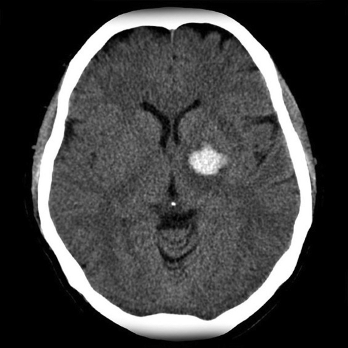
Axial noncontrast head CT scan showing hyperdensity in the globus pallidus. Basal Ganglia bleed. Source: Reproduced from Radiopaedia.org , rID:2764, case courtesy of Assoc Prof Frank Gaillard, under the Creative Commons Attribution BY-NC-SA 3.0.

Axial noncontrast head CT scan showing a large lobar hemorrhage in the frontal lobe extending into the ventricles. Source: Reproduced from Radiopaedia.org , rID:10678, case courtesy of Assoc Prof Frank Gaillard, under the Creative Commons Attribution BY-NC-SA 3.0.
References
-
- Pinto V.L., Tadi P., Adeyinka A. StatPearls Publishing; Treasure Island (FL): 2021 Jan. Increased Intracranial Pressure. [Updated 2021 Sep 29]. in: StatPearls [Internet] - PubMed
-
- Mageta M., Perry A. In: Robbins and Cotran Pathologic Basis of Disease. tenth ed. Kumar V., Abbas A.K., Aster J.C., editors. Elsevier; 2021. The central nervous system; pp. 1249–1252.
LinkOut - more resources
Full Text Sources
