. 2022 Jun;56(3):158-168.
doi: 10.1007/s13139-022-00741-x. Epub 2022 Mar 8.

Relationship Between Vitamin D Status and Brain Perfusion in Neuropsychiatric Lupus

Affiliations

Relationship Between Vitamin D Status and Brain Perfusion in Neuropsychiatric Lupus

Nasreen Sultana et al. Nucl Med Mol Imaging. 2022 Jun.

Abstract

Purpose: Neuropsychiatric manifestation of lupus (NPSLE) is related with vitamin D (vit-D) deficiency which is possibly amenable to supplementation. This study was done to explore link of serum vit-D level and clinical mini-mental state examination (MMSE) with brain perfusion SPECT (BS) in patients with NPSLE.

Methods: Patients who underwent BS with the diagnosis of NPSLE and had serum levels of vit-D and MMSE within a span of 1 month were retrospectively included. The BS DICOM data were used to generate 3D surface images of brain for visual identification of regional hypoperfusion, and the z-scores from eZIS software and then to perform voxel-based regression analysis in order to explore association between serum vit-D level and cerebral perfusion deficit using SPM8. Distribution of serum vit-D level was checked across MMSE and BS z-score using R.

Results: A total 19 patients with means ± SD age of 28.4 ± 9.2 years, having mean levels of serum vit-D of 18.7 ± 9.8 ng/ml and mean MMSE scores 24.2 ± 1.6, had undergone BS. The eZIS-derived z-score fall in the category of normal in six (31.6%), mild perfusion deficit (PD) in 10 (52.6%) and moderate PD in three (15.8%) with the means ± SD of z-score being 0.52 ± 0.2, 1.72 ± 0.2, and 2.33 ± 0.2. Voxel-based analysis revealed significant positive correlation between vit-D level and hypoperfusion in brain regions related to cognitive function (p<0.05). Serum vit-D levels were significantly lower in NPSLE patients with lower MMSE scores as well as in higher eZIS z-score (p < 0.01).

Conclusions: Our results may support the utility of vit-D supplementation in NPSLE and applicability of BS as a clinical adjunct for monitoring response to vit-D supplementation.

Keywords: Brain; Cognitive function; Neuropsychiatric systemic lupus erythematosus; SPECT; Statistical parametric mapping; Vitamin D deficiency; Voxel-based analysis.


Conflict of interest statement

Competing InterestsNasreen Sultana, Azmal Kabir Sarker, Hiroshi Matsuda, Md. Amimul Ihsan, Syed Atiqul Haq, Md. Saidul Arefin, and Sheikh Nazrul Islam declare no conflict of interest.

Figures

Fig. 1
Fig. 1
Correlation matrix of 19 brain regions of hypoperfusion with 10 clinical features. The rows and columns of the matrix have been ordered using a hierarchical clustering algorithms. The dendrograms are shown on the top and left side. The right cerebellum and the right temporal lobe were excluded in this plot because no patients showed hypoperfusion in those two regions. The regional hypoperfusions were identified by visual interpretation of eZIS-generated 3D surface view images of the brain and the clinical features were assigned by some other physicians belonging to the specialty of neurology or psychiatry available through the rheumatologists’ note. The regions were as follows: AcgR and AcgL, anterior cingulate gyrus right and left; BgnR and BgnL, basal ganglia right and left; CblR and CblL, cerebellum right (excluded) and left; IfgR and IfgL, inferior frontal gyrus right and left; MfgR and MfgL middle frontal gyrus right and left; PcgR and PcgL, posterior cingulate gyrus right and left; PcnR and PcnL, precuneus right and left; ParR and ParL, parietal gyrus right and left; SfgR and SfgL, superior frontal gyrus right and left; TemR and TemL, temporal lobe right (excluded) and left. The clinical features were as follows: Anxt, anxiety; Conf, confusion; Dprs, depression; Frgt, forgetfulness; Hach, headache; Irtb, irritability; Mood, mood disorder; Movt, movement disorder; Qoli, impaired quality of life; Spch, speech disorder
Fig. 2
Fig. 2
Brain perfusion SPECT images. a Representative 3D surface images of the brain generated by eZIS showing perfusion deficit in bilateral superior and middle frontal gyri (this patient had a Z-score of 1.25); b SPM brain surface rendering; c SPM axial slice views; and d SPM glass brain with design matrix in patients with NPSLE (n=19) showing the regional cerebral hypoperfusion in bilateral upper frontal lobes, left temporal lobe, and cingulate gyrus that correlated with serum level of vit-D (P<0.05)
Fig. 3
Fig. 3
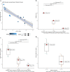
Relationship among variables; a scatterplot showing correlation between eZIS-generated BS z-score (x-axis) and serum vit-D level (y-axis) with color and size of marker corresponding to the MMSE score (lighter color and larger size indicates higher MMSE); b box plots along with jittered data points showing distribution of MMSE score across eZIS-generated z-score categories with the median MMSE scores shown on right side of each box and the p-values (Kruskal-Wallis) for pairwise comparison mentioned on top of bars between each pair of boxes; c box plots showing distribution of serum level of vit-D across eZIS-generated z-score categories with the median serum vit-D level shown on right side of each box and the p-values (Kruskal-Wallis) for pairwise comparison mentioned on top of bars between each pair of boxes; d box plots showing distribution of serum level of vit-D across MMSE score categories with the median serum vit-D level shown on right side of each box and the p-value (Mann-Whitney) for pairwise comparison is 0.001 (not displayed)

Similar articles

Cited by

References

    1. Barber MRW, Drenkard C, Falasinnu T, Hoi A, Mak A, Kow NY, et al. Global epidemiology of systemic lupus erythematosus. Nat Rev Rheumatol. 2021;17:515–532. doi: 10.1038/s41584-021-00668-1. - DOI - PMC - PubMed
    1. Islam MA, Khandker SS, Alam SS, Kotyla P, Hassan R. Vitamin D status in patients with systemic lupus erythematosus (SLE): a systematic review and meta-analysis. Autoimmun Rev. 2019;18:102392. doi: 10.1016/j.autrev.2019.102392. - DOI - PubMed
    1. Hassanalilou T, Khalili L, Ghavamzadeh S, Shokri A, Payahoo L, Bishak YK. Role of vitamin D deficiency in systemic lupus erythematosus incidence and aggravation. Auto Immun Highlights. 2017;9:1. - PMC - PubMed
    1. Tay SH, Ho CS, Ho RC, Mak A. 25-Hydroxyvitamin D3 deficiency independently predicts cognitive impairment in patients with systemic lupus erythematosus. PLoS One. 2015;10:e0144149. doi: 10.1371/journal.pone.0144149. - DOI - PMC - PubMed
    1. Cui X, Gooch H, Petty A, McGrath JJ, Eyles D. Vitamin D and the brain: genomic and non-genomic actions. Mol Cell Endocrinol. 2017;453:131–143. doi: 10.1016/j.mce.2017.05.035. - DOI - PubMed

LinkOut - more resources