. 2022 May 15;11(5):bio059206.
doi: 10.1242/bio.059206. Epub 2022 May 24.

midline represses Dpp signaling and target gene expression in Drosophila ventral leg development

Affiliations

midline represses Dpp signaling and target gene expression in Drosophila ventral leg development

Lindsay A Phillips et al. Biol Open. .

Abstract

Ventral leg patterning in Drosophila is controlled by the expression of the redundant T-box Transcription factors midline (mid) and H15. Here, we show that mid represses the Dpp-activated gene Daughters against decapentaplegic (Dad) through a consensus T-box binding element (TBE) site in the minimal enhancer, Dad13. Mutating the Dad13 DNA sequence results in an increased and broadening of Dad expression. We also demonstrate that the engrailed-homology-1 domain of Mid is critical for regulating the levels of phospho-Mad, a transducer of Dpp-signaling. However, we find that mid does not affect all Dpp-target genes as we demonstrate that brinker (brk) expression is unresponsive to mid. This study further illuminates the interplay between mechanisms involved in determination of cellular fate and the varied roles of mid.

Keywords: Dpp-signaling; Limb development; T-box transcription factors; Tissue patterning.


Conflict of interest statement

Competing interests The authors declare no competing or financial interests.

Figures

Fig. 1.
Fig. 1.
Mid blocks Dpp activation of Dad13-driven expression. (A-B) Third-instar leg discs expressing Dad13-nRFP (red, single channel) and loss-of-function H15 and mid (lack of green) clones. Clones lacking H15 and mid have (A) increased Dad13-nRFP reporter expression (white arrowhead, lower inset), or (B) expanded Dad13-nRFP reporter expression (white arrowhead, lower inset; n=7). (C) Discs with UAS-tkvQD gain-of-function clones (green) driven by AyGal4 driver showed ectopic Dad13-driven expression (red, single channel, lower inset; n=12), while (D) clones co-expressing UAS-tkvQD and UAS-mid+ (green) did not induce ectopic Dad13-driven expression (red, single channel, lower inset clone outline; n=13). All imaginal discs in this report are orientated dorsal up, anterior left.
Fig. 2.
Fig. 2.
Mid repressed Dad in part though the TBE. (A) Dad13 enhancer fragment sequences, showing the 2x SBE sequence (blue) and the TBE sequence (green). Two G to T substitutions in the TBE were generated for the Dad13TBE construct (fuchsia). (B) Dad13 expression was detected by lacZ with strong staining in the dorsal domain and weak staining in the ventral domain (arrowhead; n=37). (B′) Magnified image of ventral Dad13-lacZ reporter expression. (C,C′) UAS-mid+ gain-of-function clones (GFP) partially repressed Dad13 expression (red channel, clone outline, C′; n=8). (D) Dad13TBE-lacZ reporter expression is broader and more intense in the ventral domain relative to Dad13 (arrowhead). Dorsal expression driven by Dad13TBE is also stronger compared to Dad13 (n=35). (D′) Magnified image of ventral of Dad13TBE-lacZ reporter expression. (E,E′) Example of a UAS-mid+ gain-of-function clone (GFP) which maintains normal levels of ventral Dad13TBE-lacZ reporter expression. No repression of Dad13TBE-lacZ reporter expression was seen compared to adjacent regions outside the clone (red channel, outline, E′; n=11). (F) Fluorescent signal intensities of Dad13-lacZ and Dad13TBE-lacZ for pairs of adjacent cells inside and outside of a UAS-mid+ gain-of-function clone were measured and compared to create ratios of outside/inside. Five clones were measured for each Dad13 strain (Dad13-lacZ or Dad13TBE-lacZ) with two to five cell pairs being measured per mid clone. The overall mean ratio of cell pairs outside versus inside of a UAS-mid+ gain-of-function clone in a Dad13-lacZ leg imaginal discs was 1.95. The overall mean ratio of cell pairs outside versus inside of a UAS-mid+ gain-of-function clone in a Dad13TBE-lacZ leg imaginal discs was 1.19. Statistical analysis used the Unpaired t-test with two-tailed P-value. Bars representing the mean, s.d., and significance indicated, **** P-value≤0.0001.
Fig. 3.
Fig. 3.
Mid does not affect brk expression. (A) H15 mid loss-of-function (lack of green, arrowhead) clones have no effect on Brk expression, as detected by a Brk antibody (red, lower inset clone outline; n=41). (B) Discs expressing AyGal4 UAS-mid+ gain-of-function clones (green) do not affect brk-lacZ expression, as detected by anti-β-Galactosidase staining (red, lower inset clone outline; n=34). (C) Clones expressing UAS-tkvQD (green, top inset) have a dramatic repressive effect on brk-lacZ levels, with only residual brk-lacZ expression remaining (red, lower inset; n=7). (D) Clones co-expressing UAS-tkvQD and UAS-mid+ (green, top inset) also greatly reduce brk-lacZ expression, with only slightly higher remaining brk-lacZ expression (red, lower inset; n=7).
Fig. 4.
Fig. 4.
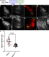
The eh1 domain is involved in Dpp repression. (A) AyGal4 gain-of-function clones expressing UAS-tkvQD result in outgrowths and dorsal transformation (arrowhead), 12.5% of legs had ectopic outgrowths under these conditions (n=181). (C) When UAS-tkvQD was co-expressed with UAS-mid+-Flag, the outgrowths were less severe (arrowhead) indicating suppression of the tkv gain-of-function phenotype. The effect was also less frequent with only 1.9% of legs having ectopic outgrowths (n=309). (E) Clones co-expressing UAS-tkvQD and UAS-mideh1-Flag had deformities (arrowhead) similar to clones expressing UAS-tkvQD alone, where 9.4% of legs had ectopic outgrowths (n=106). The clones scored in adult cuticles in panels A, C and E are not marked in these experiments but resemble the effect of marked tkvQD clones in other experiments (data not shown). pMad staining (red, greyscale single channel) was upregulated in clones (green) expressing UAS-tkvQD (B,B′,B″; n=40) and clones co-expressing UAS-tkvQD and UAS-mideh1-Flag (green) (F,F′,F″; n=24). When UAS-mid+-Flag was expressed in clones (green) along with UAS-tkvQD, pMad staining was less elevated (D,D′,D″; n=22). (G) The difference in pMad levels staining between UAS-tkvQD clones and UAS-tkvQD, UAS-mideh1-Flag was not significant. However, clones expressing UAS-tkvQD, UAS-mid+-Flag had significantly lower pMad compared to the other two conditions. The mean values were UAS-tkvQD 1.22×10⁷, UAS-tkvQD, UAS-mid+-Flag 6.73×10⁶ and UAS-tkvQD, UAS-mideh1-Flag 1.16×10⁷. Statistical analysis used the Tukey's multiple comparisons test, with bars representing the mean, s.d., and significance indicated, *P-value≤0.05 and **P-value≤0.01.

Similar articles

References

    1. Buescher, M., Svendsen, P. C., Tio, M., Miskolczi-McCallum, C., Tear, G., Brook, W. J. and Chia, W. (2004). Drosophila T box proteins break the symmetry of hedgehog-dependent activation of wingless. Curr. Biol. 14, 1694-1702. 10.1016/j.cub.2004.09.048 - DOI - PubMed
    1. Deliu, L. P., Ghosh, A. and Grewal, S. S. (2017). Investigation of protein synthesis in Drosophila larvae using puromycin labelling. Biol. Open 6, 1229-1234. 10.1242/bio.026294 - DOI - PMC - PubMed
    1. Estella, C. and Mann, R. S. (2008). Logic of Wg and Dpp induction of distal and medial fates in the Drosophila leg. Development 135, 627-636. 10.1242/dev.014670 - DOI - PMC - PubMed
    1. Formaz-Preston, A., Ryu, J.-R., Svendsen, P. C. and Brook, W. J. (2012). The Tbx20 homolog Midline represses wingless in conjunction with Groucho during the maintenance of segment polarity. Dev. Biol. 369, 319-329. 10.1016/j.ydbio.2012.07.004 - DOI - PubMed
    1. Gao, S., Steffen, J. and Laughon, A. (2005). Dpp-responsive silencers are bound by a trimeric Mad-Medea complex. J. Biol. Chem. 280, 36158-36164. 10.1074/jbc.M506882200 - DOI - PubMed

Publication types

MeSH terms

LinkOut - more resources