. 2023 Jul;86(1):282-296.
doi: 10.1007/s00248-022-02033-5. Epub 2022 May 24.

Interactions of Fungi and Algae from the Greenland Ice Sheet

Affiliations

Interactions of Fungi and Algae from the Greenland Ice Sheet

L Perini et al. Microb Ecol. 2023 Jul.

Abstract

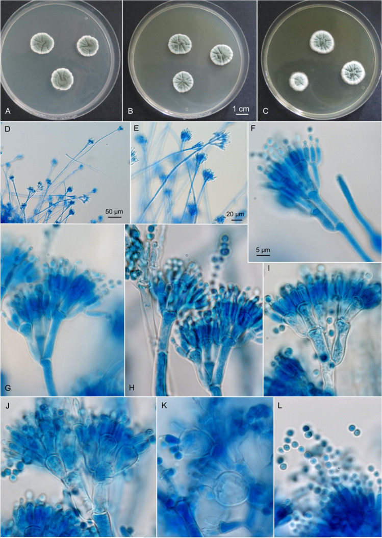
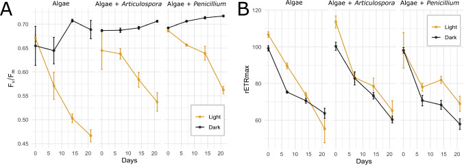
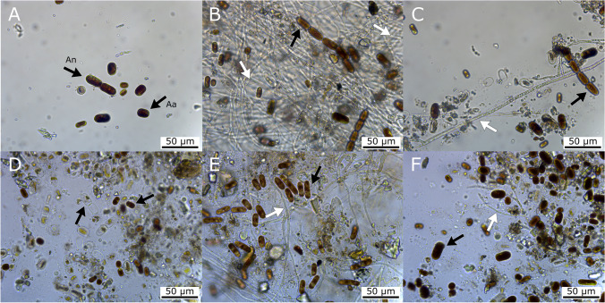
Heavily pigmented glacier ice algae Ancylonema nordenskiöldii and Ancylonema alaskanum (Zygnematophyceae, Streptophyta) reduce the bare ice albedo of the Greenland Ice Sheet, amplifying melt from the largest cryospheric contributor to eustatic sea-level rise. Little information is available about glacier ice algae interactions with other microbial communities within the surface ice environment, including fungi, which may be important for sustaining algal bloom development. To address this substantial knowledge gap and investigate the nature of algal-fungal interactions, an ex situ co-cultivation experiment with two species of fungi, recently isolated from the surface of the Greenland Ice Sheet (here proposed new species Penicillium anthracinoglaciei Perini, Frisvad and Zalar, Mycobank (MB 835602), and Articulospora sp.), and the mixed microbial community dominated by glacier ice algae was performed. The utilization of the dark pigment purpurogallin carboxylic acid-6-O-β-D-glucopyranoside (C18H18O12) by the two fungi was also evaluated in a separate experiment. P. anthracinoglaciei was capable of utilizing and converting the pigment to purpurogallin carboxylic acid, possibly using the sugar moiety as a nutrient source. Furthermore, after 3 weeks of incubation in the presence of P. anthracinoglaciei, a significantly slower decline in the maximum quantum efficiency (Fv/Fm, inverse proxy of algal stress) in glacier ice algae, compared to other treatments, was evident, suggesting a positive relationship between these species. Articulospora sp. did uptake the glycosylated purpurogallin, but did not seem to be involved in its conversion to aglycone derivative. At the end of the incubation experiments and, in conjunction with increased algal mortality, we detected a substantially increasing presence of the zoosporic fungi Chytridiomycota suggesting an important role for them as decomposers or parasites of glacier ice algae.

Keywords: Greenland Ice Sheet; HPLC; Light microscopy; Penicillium anthracinoglaciei; Purpurogallin carboxylic acid; Purpurogallin carboxylic acid-6-O-β-D-glucopyranoside; SEM.


Conflict of interest statement

The authors declare no competing interests.

Figures

Fig. 1
Fig. 1
Penicillium anthracinoglaciei sp. nov. AC Colonies grown at 25 °C for 7 days. A CYA. B MEA. C YES. DL Conidiophores and conidia on MEA. DH EXF-11443T. I EXF-10580. J EXF-10222. K EXF-10226. L EXF-11454. Scale bar indicated in F valid also for GL
Fig. 2
Fig. 2
A Glacier algae maximum quantum efficiency Fv/Fm (mean ± standard error, n = 3) during the incubation period across both light (orange) and dark (black) treatments. B rETRmax (maximum relative electron transport rate) (mean ± standard error, n = 3) over the incubation period in each treatment for light (orange) and dark (black) conditions
Fig. 3
Fig. 3
AC Light microscopy, light condition, 3-week incubation. A algal control; B glacier ice algae + Articulospora, in this treatment algae are entrapped in the mycelial network; C glacier ice algae + Penicillium anthracinoglaciei. DF Light microscopy, dark condition, 5-month incubation. D algal control; E glacier ice algae + Articulospora; F glacier ice algae + Penicillium anthracinoglaciei. In the latter treatment, the algae retained their characteristic dark pigmentation. Algal cells: black arrows (Aa, Ancylonema alaskanum; An, Ancylonema nordenskioeldii); fungal hyphae: white arrows
Fig. 4
Fig. 4
Light microscopy, light condition, 5-month incubation. Penicillium treatment. Chytrids releasing zoospores (A, black arrow) and interacting with the algae through rhizoids (white arrow) (B). Eucarpic-monocentric thalli containing zoospores (white arrowheads) on long pedicel-like rhizoids (black arrowheads) are clearly visible (C)
Fig. 5
Fig. 5
Scanning electron micrographs of algal-fungal co-cultivations. A, D Algal control (Aa, Ancylonema alaskanum; An, Ancylonema nordenskioeldii; asterisk - dead cell). Algae (black arrows) incubated with Articulospora sp. (B, E), and P. anthracinoglaciei (C, F) hyphae (white arrows) after 2 months and 5 months of incubation in light condition. B Articulospora sp. started developing a putative lichenoid structure with embedded glacier ice algae (photobiont) inside the hyphal network. DF After 5 months of incubation, cultures appeared more compact after the preparation for SEM (p, mineral particles)
Fig. 6
Fig. 6
Scanning electron micrograph of an eucarpic-monocentric thallus (white arrowhead) interacting with glacier ice algae (A) and their rhizoidal system (white arrows) branching outside of the algal substrate (B) under light condition after 5 months of incubation
Fig. 7
Fig. 7
HPLC results as monitored at 350 nm indicated the relative amounts of purpurogallin carboxylic acid-6-O-β-D-glycopyranoside and purpurogallin carboxylic acid aglycone across fungal and pigment controls and treatments after 6 weeks of incubation, both total (extracellular + intracellular) and intracellular. Values were calculated based on the chromatographic peak height and expressed in mAU (mean ± standard error, n = 3). The relative percentage on the top of the bars indicates the amount of purpurogallin glycopyranoside transformed into purpurogallin aglycone

References

    1. Procházková L, Řezanka T, Nedbalová L, Remias D. Unicellular versus filamentous: the glacial alga Ancylonema alaskana comb. et stat. nov. and its ecophysiological relatedness to Ancylonema nordenskioeldii (zygnematophyceae, streptophyta) Microorganisms. 2021;9:1–15. doi: 10.3390/microorganisms9051103. - DOI - PMC - PubMed
    1. Yallop ML, Anesio AM, Perkins RG, et al. Photophysiology and albedo-changing potential of the ice algal community on the surface of the Greenland ice sheet. ISME J. 2012;6:2302–2313. doi: 10.1038/ismej.2012.107. - DOI - PMC - PubMed
    1. Stibal M, Box JE, Cameron KA, et al. Algae drive enhanced darkening of bare ice on the Greenland ice sheet. Geophys Res Lett. 2017;44:11,463–11,471. doi: 10.1002/2017GL075958. - DOI
    1. Lutz S, McCutcheon J, McQuaid JB, Benning LG. The diversity of ice algal communities on the Greenland ice sheet as revealed by oligotyping. Microb Genomics. 2018;4:1–10. doi: 10.1099/mgen.0.000159. - DOI - PMC - PubMed
    1. Williamson CJ, Anesio AM, Cook J et al (2018) Ice algal bloom development on the surface of the Greenland ice sheet. FEMS Microbiol Ecol 94:fiy025. 10.1093/femsec/fiy025 - PMC - PubMed

LinkOut - more resources