. 2022 Sep;49(11):3651-3667.
doi: 10.1007/s00259-022-05842-5. Epub 2022 May 24.

Preclinical evaluation of FAP-2286 for fibroblast activation protein targeted radionuclide imaging and therapy

Affiliations

Preclinical evaluation of FAP-2286 for fibroblast activation protein targeted radionuclide imaging and therapy

Dirk Zboralski et al. Eur J Nucl Med Mol Imaging. 2022 Sep.

Abstract

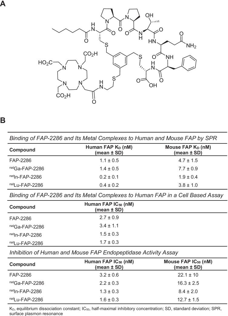
Purpose: Fibroblast activation protein (FAP) is a membrane-bound protease that has limited expression in normal adult tissues but is highly expressed in the tumor microenvironment of many solid cancers. FAP-2286 is a FAP-binding peptide coupled to a radionuclide chelator that is currently being investigated in patients as an imaging and therapeutic agent. The potency, selectivity, and efficacy of FAP-2286 were evaluated in preclinical studies.

Methods: FAP expression analysis was performed by immunohistochemistry and autoradiography on primary human cancer specimens. FAP-2286 was assessed in biochemical and cellular assays and in in vivo imaging and efficacy studies, and was further evaluated against FAPI-46, a small molecule-based FAP-targeting agent.

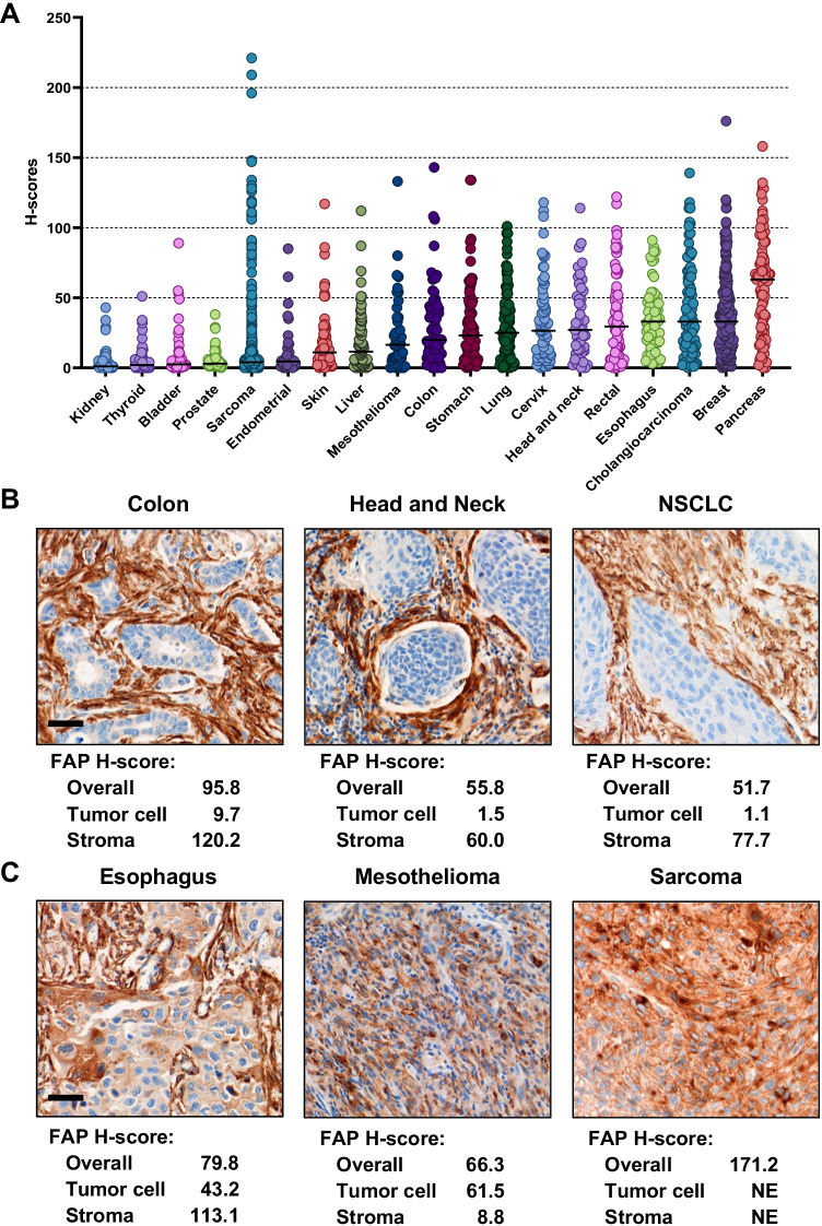
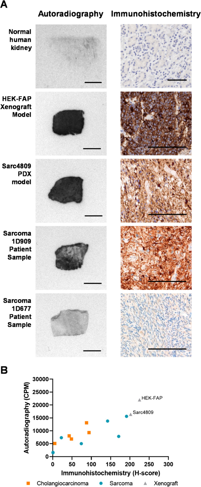
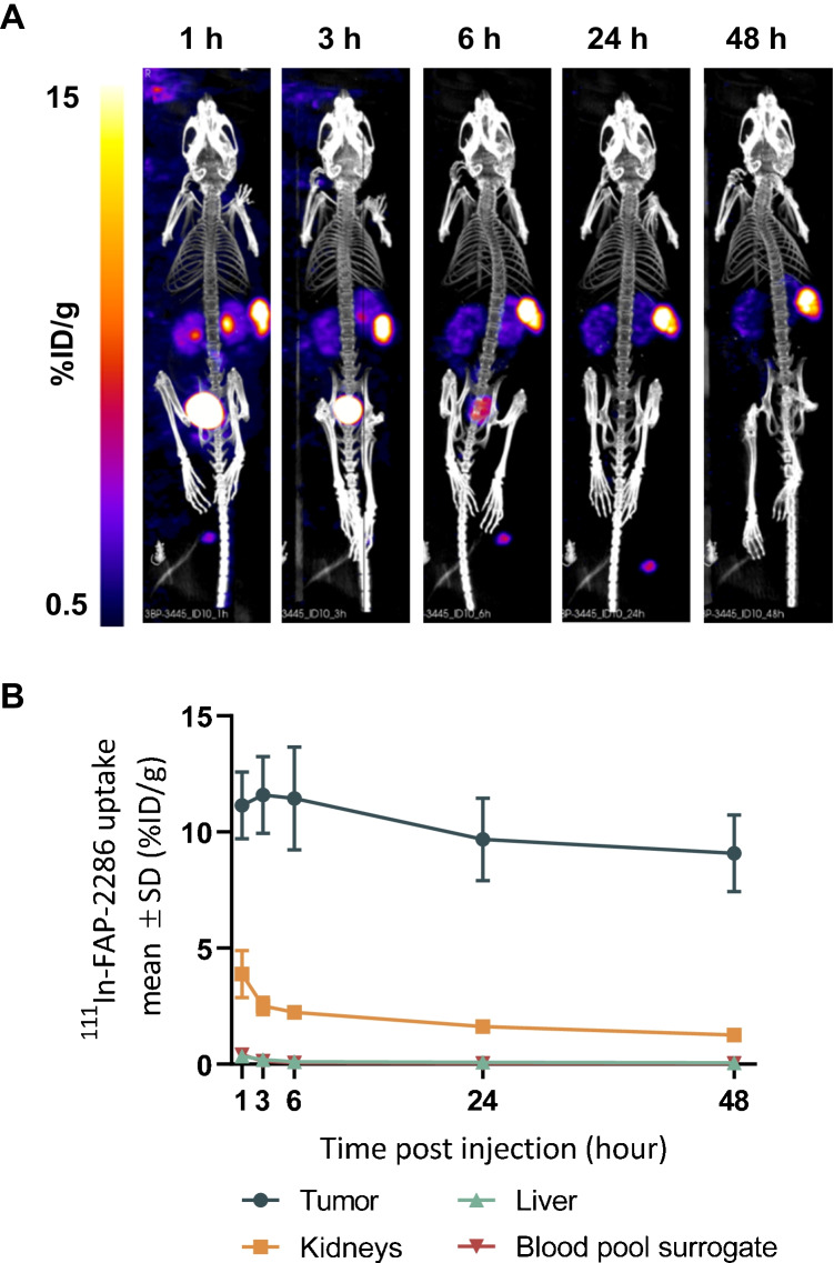
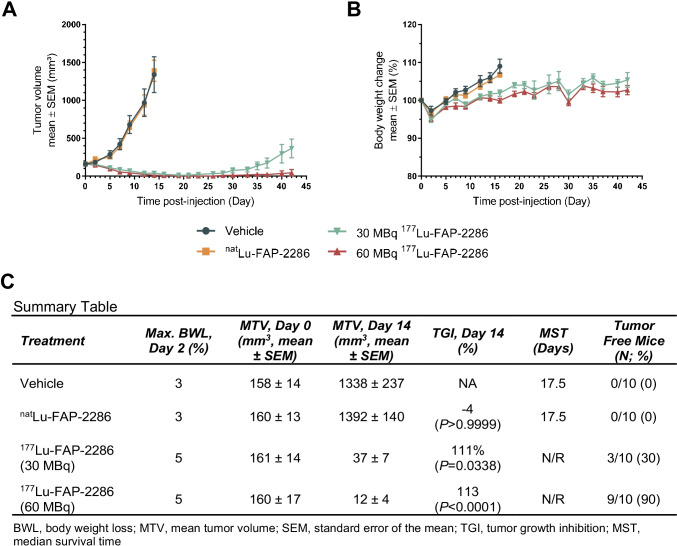
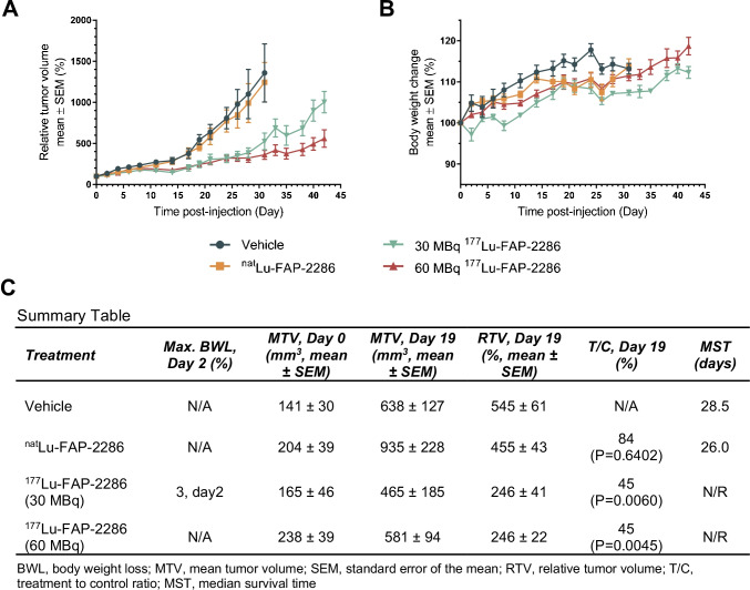
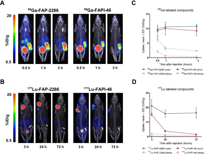
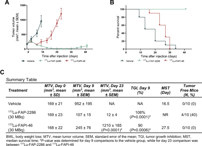
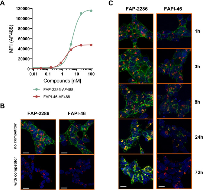
Results: Immunohistochemistry confirmed elevated levels of FAP expression in multiple tumor types including pancreatic, breast, and sarcoma, which correlated with FAP binding by FAP-2286 autoradiography. FAP-2286 and its metal complexes demonstrated high affinity to FAP recombinant protein and cell surface FAP expressed on fibroblasts. Biodistribution studies in mice showed rapid and persistent uptake of 68Ga-FAP-2286, 111In-FAP-2286, and 177Lu-FAP-2286 in FAP-positive tumors, with renal clearance and minimal uptake in normal tissues. 177Lu-FAP-2286 exhibited antitumor activity in FAP-expressing HEK293 tumors and sarcoma patient-derived xenografts, with no significant weight loss. In addition, FAP-2286 maintained longer tumor retention and suppression in comparison to FAPI-46.

Conclusion: In preclinical models, radiolabeled FAP-2286 demonstrated high tumor uptake and retention, as well as potent efficacy in FAP-positive tumors. These results support clinical development of 68Ga-FAP-2286 for imaging and 177Lu-FAP-2286 for therapeutic use in a broad spectrum of FAP-positive tumors.

Keywords: CAF; FAP; PTRT; Theranostic.


Conflict of interest statement

D. Zboralski, A. Hoehne, A. Bredenbeck, A. Schumann, E. Schneider, J. Ungewiss, M. Paschke, C. Haase, J.L. von Hacht, U. Reineke, C. Smerling, and F. Osterkamp are employees of 3B Pharmaceuticals GmbH and are named inventors of FAP-2286. M. Paschke, U. Reineke, C. Smerling, and F. Osterkamp are cofounders of 3B Pharmaceuticals GmbH. M. Nguyen, J. Lenore, T. Kwan, K.K. Lin, T.C. Harding, J. Xiao, and A.D. Simmons are or have been employees of Clovis Oncology, Inc., and may own stock or have stock options in the company.

Figures

Fig. 1
Fig. 1
FAP expression in human solid cancers. H-score quantification of FAP expression in TMA is plotted in dot plot graph by cancer types (overall; ImageScope). Median values are indicated by horizontal black bars (A). Representative images of FAP expression limited to the stroma. H-score quantification of FAP expression in the whole tumor area (overall; Visiopharm) and in the tumor cell and stroma compartment (HALO). Scale bar, 50 µm (B). Representative images of FAP expression in tumor cells. H-score quantification of FAP expression in the whole tumor area (overall; Visiopharm) and in the tumor cell and stroma compartment (HALO). NE, not evaluable; NSCLC, non-small cell lung cancer. Scale bar, 50 µm (C)
Fig. 2
Fig. 2
FAP-2286 structure (A) and in vitro characterization of FAP-2286 and its metal complexes summary table (B)
Fig. 3
Fig. 3
Correlation of FAP expression analysis by immunohistochemistry and autoradiography using 111In-FAP-2286. Representative images of patient and mouse xenograft tumors are shown by autoradiography (left, 500 µm) and immunohistochemistry (right, 100 μm) (A). FAP levels by autoradiography demonstrate correlation to immunohistochemistry in patient cholangiocarcinoma and sarcoma tumor sections (Pearson correlation coefficient r = 0.79, P = 0.002) (B)
Fig. 4
Fig. 4
Imaging of HEK-FAP tumor-bearing mice with 111In-FAP-2286. SPECT images of one representative mouse at 5 different timepoints are shown (A). Quantification of 111In-FAP-2286 uptake as mean ± SD %ID/g (n = 9) in tumor, liver, kidney, and blood pool surrogate at various timepoints after injection (B)
Fig. 5
Fig. 5
Efficacy of 177Lu-FAP-2286 treatment in the HEK-FAP tumor model. HEK-FAP tumor-bearing mice were treated with vehicle, natLu-FAP-2286, or 177Lu-FAP-2286 at 30 or 60 MBq (n = 10 mice/group). Mean tumor volumes ± SEM (A), mean body weight change ± SEM (B), and a summary table (C) are shown
Fig. 6
Fig. 6
Efficacy of 177Lu-FAP-2286 treatment in the Sarc4809 tumor model. Sarc4809 tumor-bearing mice were treated with vehicle, natLu-FAP-2286, or 177Lu-FAP-2286 at 30 or 60 MBq (n = 10 mice/group). Mean relative tumor volumes ± SEM (A), mean body weight change ± SEM (B), and a summary table (C) are shown
Fig. 7
Fig. 7
Biodistribution and tumor retention of FAP-2286 and FAPI-46 in HEK-FAP xenografts. Uptake of 68Ga-FAP-2286 and 68Ga-FAPI-46 (A) was assessed at 0.5, 1, and 3 h; and 177Lu-FAP-2286 and 177Lu-FAPI-46 (B) at 3, 24, and 72 h after dosing (n = 3 per group). One representative mouse at the 3 different timepoints is shown for FAP-2286 (left image) and FAPI-46 label radiotracers (right image). Time series plots show radiotracers accumulation as mean %ID/g ± SD at timepoints after injection in tumors and kidneys (C, D)
Fig. 8
Fig. 8
Efficacy of FAP-2286 and FAPI-46 in HEK-FAP xenografts. HEK-FAP tumor-bearing mice were treated with vehicle, 177Lu-FAP-2286, or 177Lu-FAPI-46 (30 MBq, n = 10 mice/group). Mean tumor volumes ± SEM (A) and survival curves (B) and a summary table (C) are shown
Fig. 9
Fig. 9
Cellular internalization and clearance of AF488-labeled FAP-2286 and FAPI-46 in HEK-FAP cells. HEK-FAP cells were incubated with various concentrations of AF488-labeled FAP-2286 or FAPI-46 for 1 h at 37 °C. MFI was measured by flow cytometry and plotted against compound concentration (A). HEK-FAP cells were incubated with 5 nM AF488-labeled FAP-2286 or FAPI-46 for 1 h at 37 °C (top images), and an excess of 5 µM unlabeled competitor compound was used for blocking (bottom images) (B). Cells were further incubated for an additional 1, 3, 8, 24, and 72 h (C). For visualization of lysosomal compartments, LysoTracker DeepRed was used. Cell nuclei were stained with Hoechst 33324. Images were taken with a Keyence fluorescence microscope. Scale bar 10 µm

References

    1. Scanlan MJ, Raj BK, Calvo B, Garin-Chesa P, Sanz-Moncasi MP, Healey JH, et al. Molecular cloning of fibroblast activation protein alpha, a member of the serine protease family selectively expressed in stromal fibroblasts of epithelial cancers. Proc Natl Acad Sci USA. 1994;91(12):5657–5661. doi: 10.1073/pnas.91.12.5657. - DOI - PMC - PubMed
    1. Aertgeerts K, Levin I, Shi L, Snell GP, Jennings A, Prasad GS, et al. Structural and kinetic analysis of the substrate specificity of human fibroblast activation protein alpha. J Biol Chem. 2005;280(20):19441–19444. doi: 10.1074/jbc.C500092200. - DOI - PubMed
    1. Park JE, Lenter MC, Zimmermann RN, Garin-Chesa P, Old LJ, Rettig WJ. Fibroblast activation protein, a dual specificity serine protease expressed in reactive human tumor stromal fibroblasts. J Biol Chem. 1999;274(51):36505–36512. doi: 10.1074/jbc.274.51.36505. - DOI - PubMed
    1. Waumans Y, Baerts L, Kehoe K, Lambeir AM, De Meester I. The dipeptidyl peptidase family, prolyl oligopeptidase, and prolyl carboxypeptidase in the immune system and inflammatory disease, including atherosclerosis. Front Immunol. 2015;6:387. doi: 10.3389/fimmu.2015.00387. - DOI - PMC - PubMed
    1. Niedermeyer J, Scanlan MJ, Garin-Chesa P, Daiber C, Fiebig HH, Old LJ, et al. Mouse fibroblast activation protein: molecular cloning, alternative splicing and expression in the reactive stroma of epithelial cancers. Int J Cancer. 1997;71(3):383–389. doi: 10.1002/(sici)1097-0215(19970502)71:3<383::aid-ijc14>3.0.co;2-h. - DOI - PubMed

Substances

LinkOut - more resources