Different Phenotypes Represent Advancing Stages of ABCA4-Associated Retinopathy: A Longitudinal Study of 212 Chinese Families From a Tertiary Center
- PMID: 35608843
- PMCID: PMC9150840
- DOI: 10.1167/iovs.63.5.28
Different Phenotypes Represent Advancing Stages of ABCA4-Associated Retinopathy: A Longitudinal Study of 212 Chinese Families From a Tertiary Center
Abstract
Purpose: To evaluate the nature and association of different phenotypes associated with ABCA4 mutations in Chinese.
Methods: All patients were recruited from our pediatric and genetic eye clinic. Detailed ocular phenotypes were characterized. The disease course was evaluated by long-term follow-up observation, with a focus on fundus changes. Cox regression was used to identify the factors associated with disease progression.
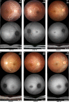
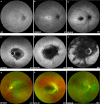
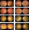
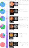
Results: A systematic review of genetic and clinical data for 228 patients and follow-up data for 42 patients indicated specific features in patients with two ABCA4 variants. Of 185 patients with available fundus images, 107 (57.8%) showed focal lesions restricted to the central macula without flecks. Among these 107 patients, 30 patients (28.0%) initially presented with relatively preserved visual acuity and inconspicuous performance on routine fundus screening. A pigmentary change in the posterior pole was observed in 22 of 185 patients (11.9%), and this change mimicked retinitis pigmentosa in 10 cases (45.5%). Follow-up visits and sibling comparisons demonstrated disease progression from cone-rod dystrophy, Stargardt disease, to retinitis pigmentosa. An earlier age of onset was associated with a more rapid decrease in visual acuity (P = 0.03). Patients with two truncation variants had an earlier age of onset.
Conclusion: Phenotypic variation in ABCA4-associated retinopathy may represent sequential changes in a single disease: early-stage Stargardt disease may resemble cone-rod dystrophy, whereas the presence of diffuse pigmentation in the late stage may mimic retinitis pigmentosa. Recognizing the natural progression of fundus changes, especially those visualized by wide-field fundus autofluorescence, is valuable for diagnostics and therapeutic decision-making.
Conflict of interest statement
Disclosure:
Figures


Similar articles
-
Full-field ERG as a predictor of the natural course of ABCA4-associated retinal degenerations.Mol Vis. 2018 Jan 4;24:1-16. eCollection 2018. Mol Vis. 2018. PMID: 29386879 Free PMC article.
-
Clinical and genetic analysis of the ABCA4 gene associated retinal dystrophy in a large Chinese cohort.Exp Eye Res. 2021 Jan;202:108389. doi: 10.1016/j.exer.2020.108389. Epub 2020 Dec 7. Exp Eye Res. 2021. PMID: 33301772
-
Genotypes Predispose Phenotypes-Clinical Features and Genetic Spectrum of ABCA4-Associated Retinal Dystrophies.Genes (Basel). 2020 Nov 27;11(12):1421. doi: 10.3390/genes11121421. Genes (Basel). 2020. PMID: 33261146 Free PMC article.
-
[From gene to disease: from the ABCA4 gene to Stargardt disease, cone-rod dystrophy and retinitis pigmentosa].Ned Tijdschr Geneeskd. 2002 Aug 24;146(34):1581-4. Ned Tijdschr Geneeskd. 2002. PMID: 12224481 Review. Dutch.
-
[Inherited retinal diseases in patients with ABCA4 gene mutations].Vestn Oftalmol. 2018;134(4):68-73. doi: 10.17116/oftalma201813404168. Vestn Oftalmol. 2018. PMID: 30166513 Review. Russian.
Cited by
-
Clinical and genetic landscape of optic atrophy in 826 families: insights from 50 nuclear genes.Brain. 2025 May 13;148(5):1604-1620. doi: 10.1093/brain/awae324. Brain. 2025. PMID: 39423307 Free PMC article.
-
Spectrum of variants associated with inherited retinal dystrophies in Northeast Mexico.BMC Ophthalmol. 2024 Feb 12;24(1):60. doi: 10.1186/s12886-023-03276-7. BMC Ophthalmol. 2024. PMID: 38347443 Free PMC article.
-
ABCA4 Deep Intronic Variants Contributed to Nearly Half of Unsolved Stargardt Cases With a Milder Phenotype.Invest Ophthalmol Vis Sci. 2025 Jan 2;66(1):65. doi: 10.1167/iovs.66.1.65. Invest Ophthalmol Vis Sci. 2025. PMID: 39883546 Free PMC article.
-
FDXR-Associated Oculopathy: Congenital Amaurosis and Early-Onset Severe Retinal Dystrophy as Common Presenting Features in a Chinese Population.Genes (Basel). 2023 Apr 21;14(4):952. doi: 10.3390/genes14040952. Genes (Basel). 2023. PMID: 37107710 Free PMC article.
-
Natural History of Stargardt Disease: The Longest Follow-Up Cohort Study.Genes (Basel). 2023 Jul 2;14(7):1394. doi: 10.3390/genes14071394. Genes (Basel). 2023. PMID: 37510299 Free PMC article.
References
-
- Allikmets R. A photoreceptor cell-specific ATP-binding transporter gene (ABCR) is mutated in recessive Stargardt macular dystrophy. Nat Genet. 1997; 17: 122. - PubMed
-
- Nasonkin I, Illing M, Koehler MR, Schmid M, Molday RS, Weber BH.. Mapping of the rod photoreceptor ABC transporter (ABCR) to 1p21-p22.1 and identification of novel mutations in Stargardt's disease. Hum Genet. 1998; 102: 21–26. - PubMed
-
- Cornelis SS, Bax NM, Zernant J, et al. .. In silico functional meta-analysis of 5,962 ABCA4 variants in 3,928 retinal dystrophy cases. Hum Mutat. 2017; 38: 400–408. - PubMed
Publication types
MeSH terms
Substances
LinkOut - more resources
Full Text Sources

