. 2022 Jun 15;132(12):e154317.
doi: 10.1172/JCI154317.

The neuronal tyrosine kinase receptor ligand ALKAL2 mediates persistent pain

Affiliations

The neuronal tyrosine kinase receptor ligand ALKAL2 mediates persistent pain

Manon Defaye et al. J Clin Invest. .

Abstract

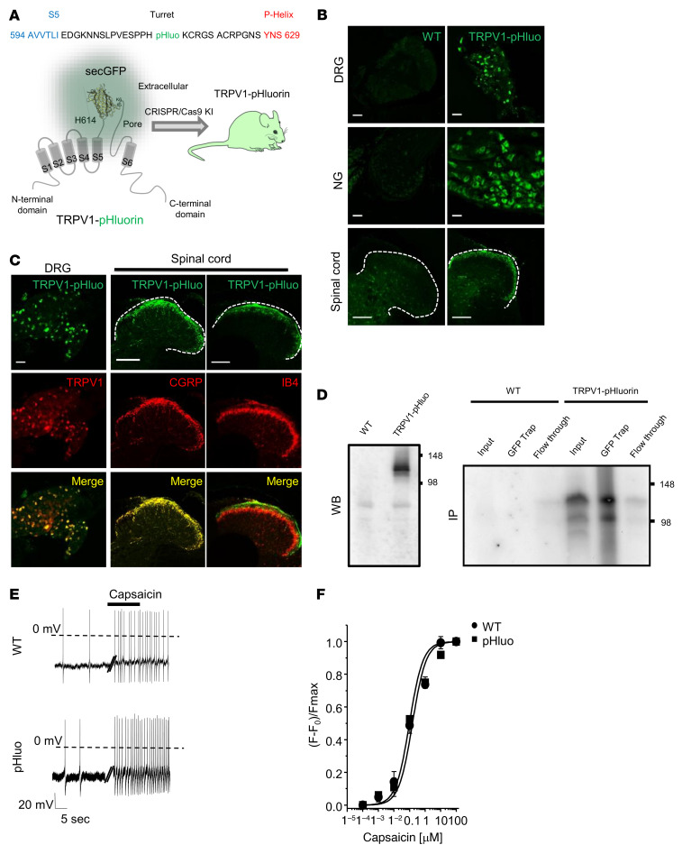
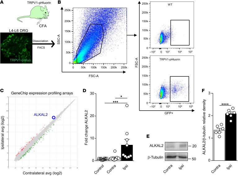
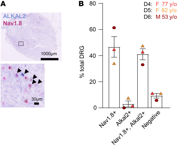
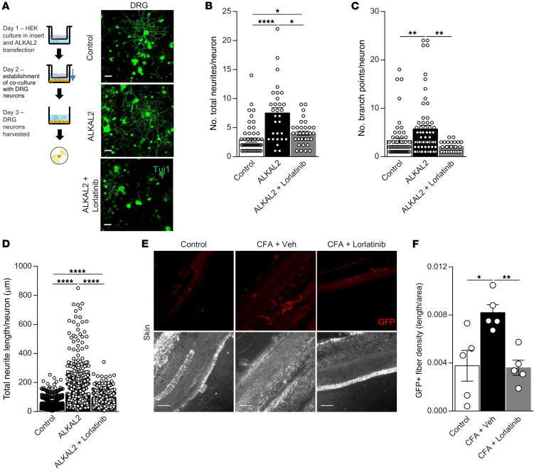
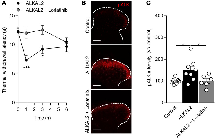
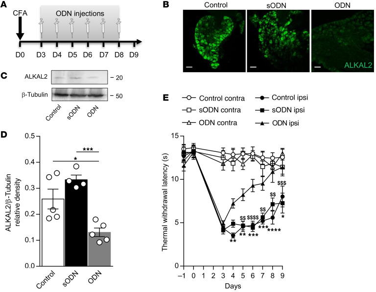
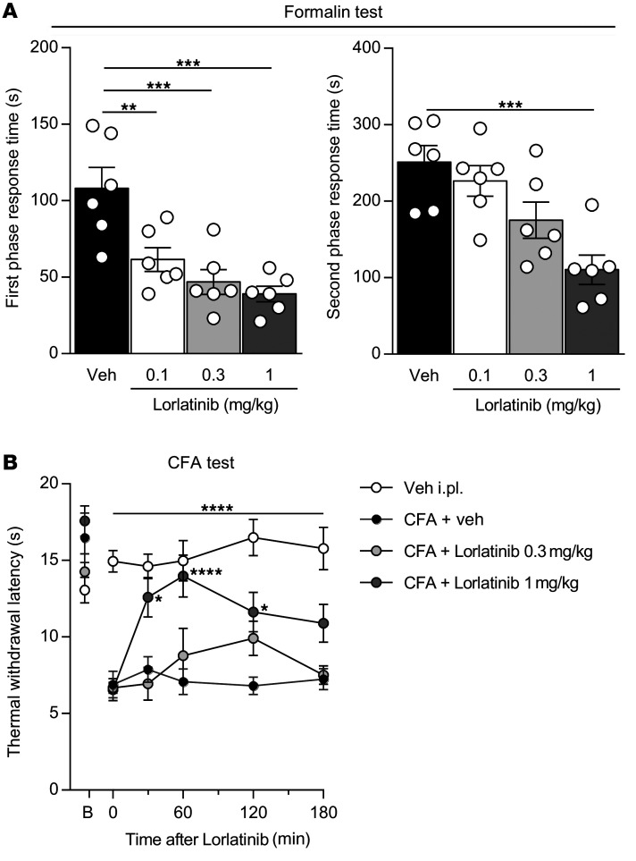
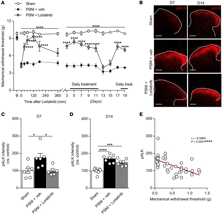
The anaplastic lymphoma kinase (ALK) is a receptor tyrosine kinase known for its oncogenic potential that is involved in the development of the peripheral and central nervous system. ALK receptor ligands ALKAL1 and ALKAL2 were recently found to promote neuronal differentiation and survival. Here, we show that inflammation or injury enhanced ALKAL2 expression in a subset of TRPV1+ sensory neurons. Notably, ALKAL2 was particularly enriched in both mouse and human peptidergic nociceptors, yet weakly expressed in nonpeptidergic, large-diameter myelinated neurons or in the brain. Using a coculture expression system, we found that nociceptors exposed to ALKAL2 exhibited heightened excitability and neurite outgrowth. Intraplantar CFA or intrathecal infusion of recombinant ALKAL2 led to ALK phosphorylation in the lumbar dorsal horn of the spinal cord. Finally, depletion of ALKAL2 in dorsal root ganglia or blocking ALK with clinically available compounds crizotinib or lorlatinib reversed thermal hyperalgesia and mechanical allodynia induced by inflammation or nerve injury, respectively. Overall, our work uncovers the ALKAL2/ALK signaling axis as a central regulator of nociceptor-induced sensitization. We propose that clinically approved ALK inhibitors used for non-small cell lung cancer and neuroblastomas could be repurposed to treat persistent pain conditions.

Keywords: Cancer; Neuroscience; Pain; Signal transduction.


Figures

Figure 1
Figure 1. Characterization of the TRPV1-pHluorin knockin mouse.
(A) PHluorin was inserted into the turret region of the TRPV1 channel, between residues H614 and K615 using CRISPR/Cas9 technology (31, 32). (B) Representative confocal images showing GFP expression in DRG, nodose ganglion (NG), and spinal cords of WT and TRPV1-pHluorin mouse. Scale bars: 50 μm (DRG and nodose ganglion); 100 μm (spinal cord). (C) Coimmunostaining of GFP and TRPV1 in DRG; coimmunostaining of GFP with markers of peptidergic (CGRP) or nonpeptidergic (IB4) neurons in the spinal dorsal horn. Scale bars: 50 μm (DRG); 100 μm (spinal cord). (D) Western blot of DRG lysates from WT and TRPV1-pHluorin mice using an anti-GFP antibody; note the specific band at approximately 125 kDa only in the transgenic mice. Immunoprecipitation of TRPV1-pHluorin from DRG lysates using GFP-trap. Membranes were then probed with an anti-TRPV1 antibody. Note the absence of band in WT animals. (E) Representative whole-cell current-clamp recordings of the capsaicin-evoked AP discharge in DRG neurons from WT and TRPV1-pHluorin mice. (F) Dose-response curve evoked by capsaicin, measured by calcium imaging on DRG neurons from WT (circles) (EC50 = 0.91 ± 0.12 μM, n = 34) or TRPV1-pHluorin (pHluo) mice (squares) (EC50 = 1.06 ± 0.09 μM, n = 28) at RT (22°C). Data are represented as mean ± SEM.
Figure 2
Figure 2. CFA inflammation induces ALKAL2 upregulation in TRPV1 neurons.
(A) Experimental approach used to conduct the microarray analysis from TRPV1 neurons, 72 hours after i.pl. CFA. (B) FACS isolation of TRPV1-pHluorin neurons. Representative FACS plot of GFP+ population in WT (top) and TRPV1-pHluorin (bottom) mice. SSC-A side scatter area; FSCA- forward scatter area. (C) Scatter plot representation of genes regulated in CFA conditions. Genes that passed a threshold of log2 fold change in differential expression analysis are represented as green when downregulated and red when upregulated. All genes are listed in Supplemental Table 1. (D) qRT-PCR assessment of ALKAL2 upregulation in the DRG ipsilateral to the CFA injection (Ipsi) (n = 9), compared with the contralateral side (Contra) (n = 9) and naive control (n = 8). Statistical analysis was performed using Kruskal-Wallis followed by Dunn’s post hoc test. *P < 0.05; ***P < 0.001. (E) Representative Western blot of ALKAL2 in the DRG ipsilateral to the CFA injection compared with the contralateral side. (F) Quantification of ALKAL2 protein level from Western blot experiments. Each dot represents a sample collected from a different animal (n = 6 per group). Statistical analysis was performed by unpaired t test (F). ****P < 0.0001. Data are represented as mean ± SEM.
Figure 3
Figure 3. Characterization of mouse ALKAL2 expression.
(A) Heatmap of the expression of ALKAL2 and selected population markers on the 17 populations of sensory neurons from DRG described in Zeisel et al. (35). (B) Representative confocal images of coimmunostaining for ALKAL2 and TRPV1, IB4, GFRα3, and NF200 in DRG neurons. Scale bars: 50 μm. (C) Dot plot summarizing the results in B: 77.64% ± 2.95% of TRPV1, 77.64% ± 3.06% of GFRα3, 41.65% ± 7.08% of IB4, and 36.46% ± 2.64% of NF200-positive neurons express ALKAL2 (each symbol represents a DRG section from n = 4 individual animals). Statistical analysis was performed using 1-way ANOVA followed by Tukey’s post hoc test. ****P < 0.001. (D) Representative confocal images of coimmunostaining for ALKAL2 and NF200, IB4, and GFRα3 in the sciatic nerve. Scale bars: 50 μm. Data are represented as mean ± SEM.
Figure 4
Figure 4. ALKAL2 expression in human DRGs.
(A) Representative RNAScope image showing expression of ALKAL2 (light blue) in human DRG neurons coexpressing Nav1.8 (pink). (B) Bar graph summarizing the results (each symbol represents an individual patient, n = 3). Data are represented as mean ± SEM.
Figure 5
Figure 5. ALKAL2 induces sprouting of DRG neurons.
(A) Schematic illustrating the coculture system used to chronically expose DRG neurons to ALKAL2. HEK cells were plated into the upper chamber of a Transwell and then transfected with ALKAL2 plasmid for 16 hours. Cells were washed, and DRG neurons were plated in the lower chamber of the Transwell for another 16 hours of coculture and then immunostained for Tuj1. Scale bars: 50 μm. ALKAL2 induces a significant increase of total neurites (B), number of branch points per neuron (C), and total neurite length per neuron (D) (<20 μm). Control, n = 19; ALKAL2, n = 12; ALKAL2+lorlatinib, n = 18. Statistical analysis was performed using Kruskal-Wallis followed by Dunn’s post hoc test. *P < 0.05; **P < 0.01; ****P < 0.001. (E) Representative confocal images illustrating the TRPV1-GFP innervation of the skin paw following i.pl. injection of CFA (3 days). Scale bars: 50 μm. (F) CFA-induced sprouting is reversed by daily administration of lorlatinib (1 mg/kg). Control, n = 5; CFA+vehicle, n = 5; CFA+lorlatinib, n = 5. Statistical analysis was performed using 1-way ANOVA followed by Tukey’s post hoc test. *P < 0.05; **P < 0.01. Data are represented as mean ± SEM.
Figure 6
Figure 6. ALKAL2 induces hyperexcitability of small DRG neurons.
(A) Resting membrane potential (RMP) of small DRG neurons in control (–56.06 ± 1.03 mV, n = 19), ALKAL2 (–55.15 ± 2.21 mV, n = 13), or ALKAL2+lorlatinib (–54.5 ± 1.53 mV, n = 13) groups. Statistical analysis was performed using 1-way ANOVA followed by Tukey’s post hoc test. (B) AP threshold in control (–35.59 ± 2.26 mV, n = 16), ALKAL2 (–40.57 ± 2.88 mV, n = 11), and ALKAL2+lorlatinib (–31.59 ± 1.76 mV, n = 9) groups. Statistical analysis was performed using 1-way ANOVA followed by Tukey’s post hoc test. (C) Representative AP discharge evoked by 100, 200, 300, and 400 pA current injections (1 s) in control, ALKAL2- (10 nM), and ALKAL2+lorlatinib-treated (10 nM+1 μM) DRG neurons. (D) Measure of AP frequency evoked by current injection in the different groups represented in E (control, n = 13; ALKAL2 [10 nM], n = 11; ALKAL2+lorlatinib treated [10 nM+1 μM], n = 9). Statistical analysis was performed using 2-way ANOVA followed by Tukey’s post hoc test. *P < 0.05; ***P < 0.001; ****P < 0.0001 versus control. $P < 0.05; $$$$P < 0.0001 versus ALKAL2+lorlatinib. Data are represented as mean ± SEM.
Figure 7
Figure 7. ALKAL2 induces pALK in the dorsal horn of the spinal cord.
(A) Administration i.t. of ALKAL2 (1 μM) promotes thermal hyperalgesia, which is reversed by the administration of lorlatinib (ALKAL2, n = 9; ALKAL2+lorlatinib, n = 8). Statistical analysis was performed using 2-way ANOVA followed by Bonferroni’s post hoc test. *P < 0.05; ***P < 0.001. (B) Representative confocal images illustrating pALK induction in the lamina I and II of the spinal dorsal horn following i.t. infusion of ALKAL2. Activation of pALK is reversed by administration of lorlatinib prior to ALKAL2 administration. Scale bars: 100 μm. (C) Bar graph of the pALK signal intensity represented in B in the spinal dorsal horn (control, n = 7; ALKAL2, n = 8; ALKAL2+lorlatinib, n = 6; 1 hour). Statistical analysis was performed using 1-way ANOVA followed by Tukey’s post hoc test. *P < 0.05. Data are represented as mean ± SEM.
Figure 8
Figure 8. ALKAL2 gene depletion reverses inflammatory pain.
(A) Schematic illustrating the experimental protocol of ALKAL2 oligodeoxynucleotide injection in the CFA pain model. (B) Representative confocal images of ALKAL2 immunostaining in DRG sections from control, ALKAL2, or scrambled ODN–treated animals. Scale bars: 50 μm. (C) Western blot of ALKAL2 in lumbar DRG lysates at D9 following injection of ALKAL2 or scrambled ODN, compared with naive control mice. (D) Bar graph illustrating the reduction in ALKAL2 protein expression in the ODN-treated animals (n = 4–6 mice per group). Statistical analysis was performed using 1-way ANOVA followed by Tukey’s post hoc test. *P < 0.05; ***P < 0.001. (E) Measure of thermal withdrawal latency in contralateral and ipsilateral hind paws of CFA-injected animals that received saline control (n = 8), scrambled (n = 7), or ALKAL2 ODN (n = 8). Statistical analysis was performed using 2-way ANOVA followed by Tukey’s post hoc test. *P < 0.05, **P < 0.01, ***P < 0.001 ****P < 0.0001 ODN ipsi vs Control ipsi; $$P< 0.01, $$$P < 0.001, $$$$P < 0.0001 ODN ipsi vs sODN ipsi. Data are represented as mean ± SEM.
Figure 9
Figure 9. Pharmacological inhibition of ALK receptor induces antinociception in inflammatory pain models.
(A) Shortening of the nociceptive behavior duration produced by i.pl. formalin (20 μl of 1.25% solution) after administration of increasing doses of lorlatinib by gavage; vehicle was administered as control (n = 6 per condition). Statistical analysis was performed using 1-way ANOVA followed by Dunnett’s post hoc test. **P < 0.01; ***P < 0.001 versus vehicle. (B) Paw withdrawal latency in response to a thermal stimulus in the CFA model showed the antinociceptive effect of lorlatinib (PBS, n = 10; CFA+vehicle, n = 10; CFA+lorlatinib, 0.3 mg/kg, n = 12; CFA+lorlatinib, 1 mg/kg, n = 10). Statistical analysis was performed using 2-way ANOVA followed by Tukey’s post hoc test. *P < 0.05; ****P < 0.0001 versus CFA+vehicle. Data are represented as mean ± SEM.
Figure 10
Figure 10. Pharmacological inhibition of ALK receptor induces antinociception in neuropathic pain models.
(A) PWT to mechanical stimuli was evaluated for 19 days after nerve injury. Lorlatinib inhibited PWT when compared with vehicle only (sham, n = 10; PSNI+vehicle, n = 10; PSNI+lorlatinib, n = 10). Statistical analysis was performed using 2-way ANOVA followed by Tukey’s post hoc test. *P < 0.05; ****P < 0.0001 versus PSNI+vehicle. (B) Representative confocal images illustrating the pALK induction in the lamina I and II of the spinal dorsal horn following PSNI. Activation of pALK is normalized by administration of lorlatinib at day 7 and reversed after stopping lorlatinib treatment at day 14. Scale bars: 100 μm. (C and D) Bar graphs of the pALK signal intensity represented in B in the spinal dorsal horn at day 7 (C) and day 14 (D) (sham, n = 7; PSNI+vehicle, n = 6–7; PSNI+lorlatinib, n = 6–7). Statistical analysis was performed using 1-way ANOVA followed by Tukey’s post hoc test.*P < 0.05; ***P < 0.001; ****P < 0.0001. (E) Negative correlation between activation of ALK and PWT. Statistical analysis was performed using Pearson’s test. ****P < 0.0001. Data are represented as mean ± SEM.

References

    1. Lapointe TK, et al. TRPV1 sensitization mediates postinflammatory visceral pain following acute colitis. Am J Physiol Gastrointest Liver Physiol. 2015;309(2):G87–G99. doi: 10.1152/ajpgi.00421.2014. - DOI - PubMed
    1. Bourinet E, et al. Calcium-permeable ion channels in pain signaling. Physiol Rev. 2014;94(1):81–140. doi: 10.1152/physrev.00023.2013. - DOI - PubMed
    1. Basbaum AI, et al. Cellular and molecular mechanisms of pain. Cell. 2009;139(2):267–284. doi: 10.1016/j.cell.2009.09.028. - DOI - PMC - PubMed
    1. Tominaga M, et al. The cloned capsaicin receptor integrates multiple pain-producing stimuli. Neuron. 1998;21(3):531–543. doi: 10.1016/S0896-6273(00)80564-4. - DOI - PubMed
    1. Coleridge JC, Coleridge HM. Afferent vagal C fibre innervation of the lungs and airways and its functional significance. Rev Physiol Biochem Pharmacol. 1984;99:1–110. - PubMed

Publication types

Grants and funding