. 2022 Aug;27(8):712-723.
doi: 10.1111/nep.14066. Epub 2022 Jul 5.

Peptide 17 alleviates early hypertensive renal injury by regulating the Hippo/YAP signalling pathway

Affiliations

Peptide 17 alleviates early hypertensive renal injury by regulating the Hippo/YAP signalling pathway

San-Bin Xu et al. Nephrology (Carlton). 2022 Aug.

Abstract

Aim: Hypertensive nephropathy is embodied by kidney tissue fibrosis and glomerular sclerosis, as well as renal inflammation. The Hippo/YAP (yes-associated protein, YAP) axis has been reported to promote inflammation and fibrosis and may participate in the pathogenesis of heart, vascular and renal injuries. However, the role of the Hippo/YAP pathway in hypertensive renal injury has not been reported so far. We explored the role of the Hippo/YAP signalling pathway in hypertensive renal injury and the effect of peptide 17 on its effects.

Methods: Histopathological analyses were performed based on the Masson and Haematoxylin/eosin (HE) staining approaches. Biochemical indexes were determined and immunofluorescence and western blotting were used to detect protein expression levels. The mRNA expression levels were determined by qRT-PCR.

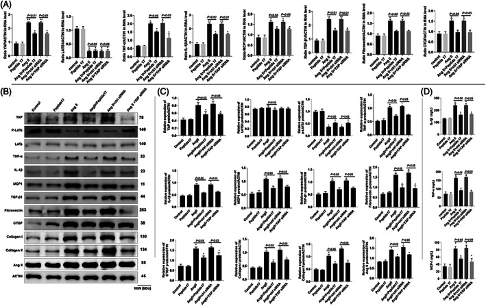
Results: Our results showed that peptide 17 reduced the systolic blood pressure (SBP) and urine protein/creatinine ratio in hypertensive rats. In addition, peptide 17 reduced the histopathological damage of kidneys in spontaneously hypertensive rats (SHRs). Moreover, peptide 17 downregulated genes in the Hippo/Yap pathway in kidney tissue of SHRs and Ang II-treated kidney cells. The expression levels of inflammatory factors TNF-α, IL-1β and MCP-1 and the pro-fibrotic factors TGF-β1, fibronectin, and CTGF were increased in the kidney of hypertensive rats, but reversed by peptide 17 treatment. Silencing of YAP had effect similar to that of peptide 17 in vivo and in vitro.

Conclusion: Peptide 17 alleviates early renal injury in hypertension by regulating the Hippo/YAP signalling pathway. These findings may be useful in the treatment of hypertensive renal injury.

Keywords: angiotensin II; hippo/YAP pathway; hypertension; peptide 17; renal damage.


Conflict of interest statement

The authors declare that they have no conflict of interest.

Figures

FIGURE 1
FIGURE 1
Peptide 17 inhibits the interaction of YAP/TEAD1 in the renal cells. The effect of peptide 17 on the interaction of YAP/TEAD1 was analysed by co‐immunoprecipitation. (A) Inhibition of the binding of myc‐TEAD1 to affinity‐purified GFP–YAP by peptide 17. (B) Inhibition of the binding of GFP–YAP to immunoprecipitated myc‐TEAD1 by Peptide 17. The experiments were repeated thrice
FIGURE 2
FIGURE 2
Peptide 17 reduces SBP and modulates biochemical parameters of kidney injury in hypertensive rats. (A) Body weight. (B) Ratio of kidney weight/body weight. (C) Creatine content in plasma. (D) NGAL in plasma. (E) Urine volume. (F) Albumin content in urine. (G) Albumin content in urine. (H) NGAL in urine. (I) SBP in different groups. The one‐way ANOVA followed by Tukey's multiple comparison posttests was performed. For A‐H, *p < .05 compared with WKY group; the significance difference for other group comparisons is reported in the figure panels. For I, * in orange indicates p < .05 in the comparison of SHR + ctrl siRNA with WKY, * in red indicates p < .05 in the comparison of SHR with WKY, * in green indicates p < .05 in the comparison of SHR+ peptide 17 with SHR, * in black indicates p < .05 in the comparison of SHR+ YAP siRNA with SHR. The results were obtained from six rats/group
FIGURE 3
FIGURE 3
Effect of peptide 17 on the histopathological damages of kidney in kidney tissue of rats. (A) Representative images of H&E staining. (B) Tissue damage rate from HE staining. (C) Representative images of H&E staining. (D) Masson‐positive staining areas. *p < .05, **p < .01 compared to the WKY group; the significance of the difference among the other compared groups is indicated in the figure. The results were obtained from six rats/group
FIGURE 4
FIGURE 4
Immunofluoresence analysis of YAP and heparanase in kidney tissue. (A) Representative images of immunofluorescence experimental results. (B) Ratio of immunofluorescence intensity of YAP in different groups over WKY group. (C) Ratio of immunofluorescence intensity of heparanase in different groups over WKY group. **p < .01 compared to the WKY group; the significance of the difference among the other compared groups is indicated in the figure. The results were obtained from six rats/group
FIGURE 5
FIGURE 5
Peptide 17 regulates Hippo/YAP signalling, inflammatory markers. Fibrosis markers and Ang II in kidney tissue of SHRs. (A) mRNA levels of YAP, LATS1, TNF‐α, MPC1, TGF‐β1, IL‐1β, Fibronectin, and CTGF in the kidney tissue of rats from different groups. (B) Bands of western blot analysis of protein expression. (C) Densitometry analysis of bands from the western blot analysis showing quantitative representation of gene expression. (D) Determination of Ang II content in the kidney tissue of different rat groups. *p < .05, **p < .01 compared with the WKY group; the significance of the comparisons between other groups are also indicated in the figure. The results were obtained from six rats/group
FIGURE 6
FIGURE 6
Immunofluorescence analysis of the expression of YAP in NRK‐52E cells subjected to different treatments. (A) Immuno‐stained cell photomicrographs; (B) YAP immunofluorescence intensity in cell. *p < .05, **p < .01 compared with the Control group; the significance of the comparisons between other groups are also indicated in the figure. The results were obtained from six repeated experiments
FIGURE 7
FIGURE 7
Peptide 17 regulates Hippo/YAP signalling, inflammatory markers. Fibrosis markers and Ang II in NRK‐52E cells. (A) mRNA levels of YAP, LATS1, TNF‐α, MPC1, TGF‐β1, IL‐1β, Fibronectin, and CTGF in NRK‐52E cells from different treatment groups. (B) Bands of western blot analysis of protein expression. (C) Densitometry analysis of bands from the western blot analysis showing quantitative representation of gene expression. (D) Determination of MCP‐1, TNF‐α, and IL‐1β by ELISA. *p < .05, **p < .01 compared with the control group; the significance of the comparisons between other groups are also indicated in the figure. The results were obtained from six repeated experiments

Similar articles

Cited by

References

    1. Ruiz‐Ortega M, Rayego‐Mateos S, Lamas S, Ortiz A, Rodrigues‐Diez RR. Targeting the progression of chronic kidney disease. Nat Rev Nephrol. 2020;16:269‐288. - PubMed
    1. Hamrahian SM, Falkner B. Hypertension in chronic kidney disease. Adv Exp Med Biol. 2017;956:307‐325. - PubMed
    1. Hirsch S, Hirsch J, Bhatt U, Rovin BH. Tolerating increases in the serum creatinine following aggressive treatment of chronic kidney disease, hypertension and proteinuria: pre‐renal success. Am J Nephrol. 2012;36:430‐437. - PubMed
    1. Udani S, Lazich I, Bakris GL. Epidemiology of hypertensive kidney disease. Nat Rev Nephrol. 2011;7:11‐21. - PubMed
    1. Hall JE, Granger JP, do Carmo JM, et al. Hypertension: physiology and pathophysiology. Comprehensive Phys Ther. 2012;2:2393‐2442. - PubMed