. 2022 Aug;67(2):188-200.
doi: 10.1165/rcmb.2021-0554OC.

Aberrant Multiciliogenesis in Idiopathic Pulmonary Fibrosis

Affiliations

Aberrant Multiciliogenesis in Idiopathic Pulmonary Fibrosis

Eunjoo Kim et al. Am J Respir Cell Mol Biol. 2022 Aug.

Abstract

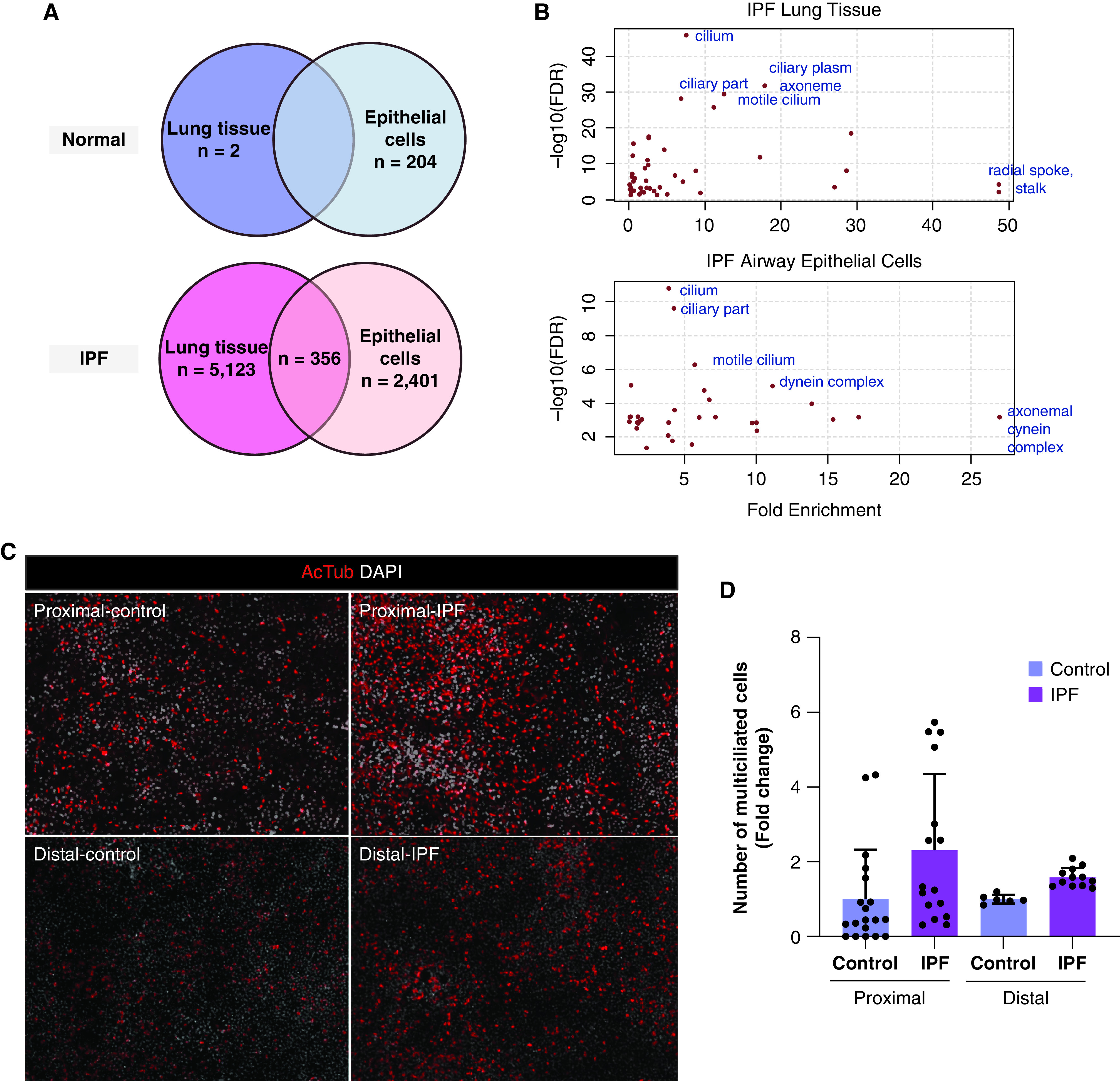
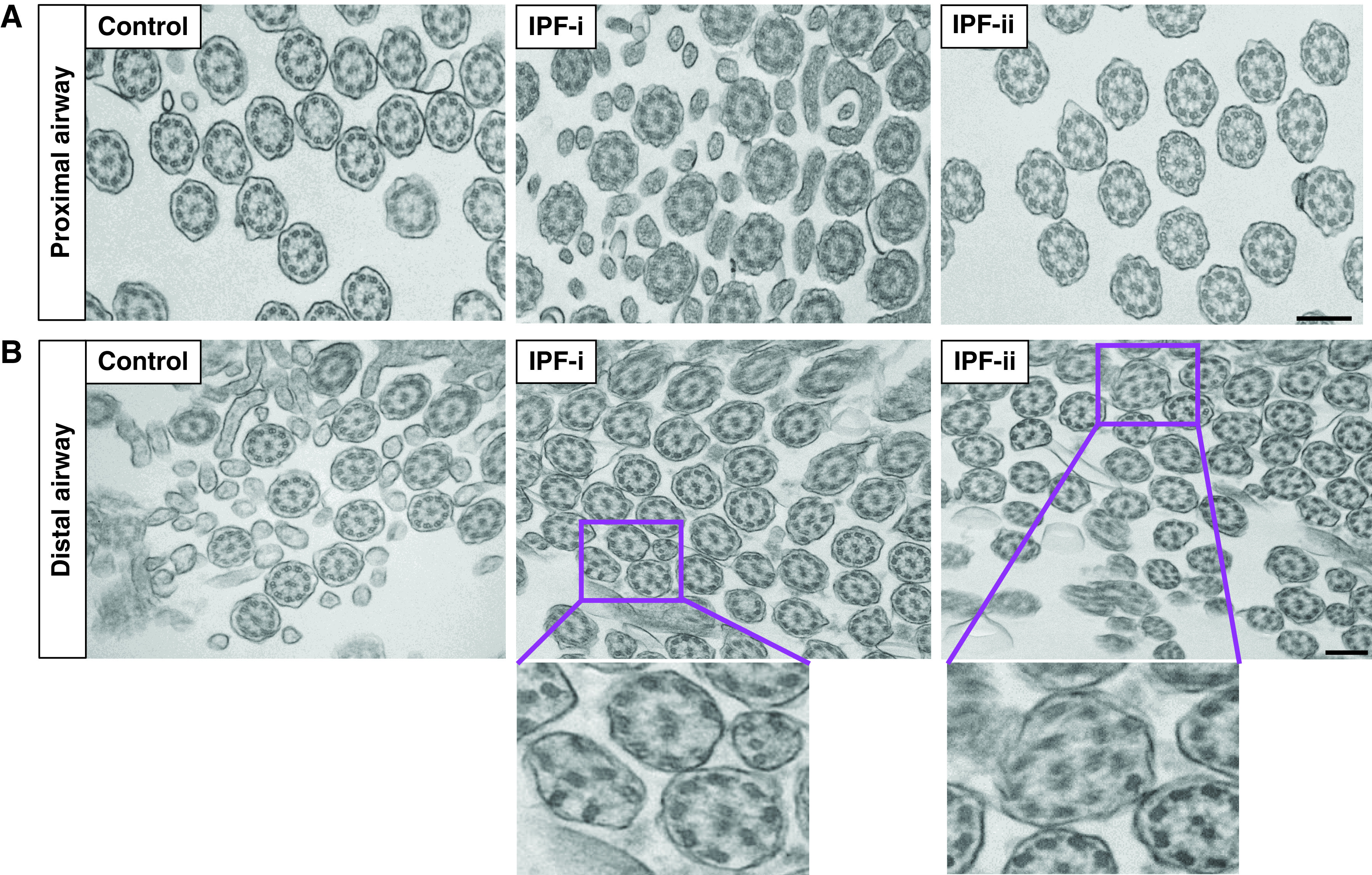
We previously identified a novel molecular subtype of idiopathic pulmonary fibrosis (IPF) defined by increased expression of cilium-associated genes, airway mucin gene MUC5B, and KRT5 marker of basal cell airway progenitors. Here we show the association of MUC5B and cilia gene expression in human IPF airway epithelial cells, providing further rationale for examining the role of cilium genes in the pathogenesis of IPF. We demonstrate increased multiciliogenesis and changes in motile cilia structure of multiciliated cells both in IPF and bleomycin lung fibrosis models. Importantly, conditional deletion of a cilium gene, Ift88 (intraflagellar transport 88), in Krt5 basal cells reduces Krt5 pod formation and lung fibrosis, whereas no changes are observed in Ift88 conditional deletion in club cell progenitors. Our findings indicate that aberrant injury-activated primary ciliogenesis and Hedgehog signaling may play a causative role in Krt5 pod formation, which leads to aberrant multiciliogenesis and lung fibrosis. This implies that modulating cilium gene expression in Krt5 cell progenitors is a potential therapeutic target for IPF.

Keywords: Ift88; Krt5 cell; abnormal structure of multiciliated cells; injury-induced multiciliogenesis.


Figures

Figure 1.
Figure 1.
Multiciliated cells are increased in idiopathic pulmonary fibrosis (IPF). (A) Summary of genes whose expression is significantly associated (false discovery rate < 0.05) with MUC5B gene expression in proximal airway epithelia and whole lung tissue of subjects with IPF and nondiseased control subjects. (B) Pathway enrichment using gene ontology categories for cellular components. (C) Acetylated tubulin (red) and DAPI (white) expression at 28 days after establishing ALI culture using control and IPF airway epithelia. (D) Fold changes in the number of multiciliated cells from ALI cultures in panel (C). Data are presented as dot plots with means ± SD (n = 3 donors per group). Statistical significance was determined using a one-way ANOVA. ALI = air–liquid interface; DAPI = 4′,6-diamidino-2-phenylindole; FDR = false discovery rate.
Figure 2.
Figure 2.
Motile cilia structure is disrupted in the IPF distal airway. Transition electron microscopy of axoneme of motile cilia of (A) proximal airway and (B) distal airway in healthy control subjects and patients with IPF. IPF-i and IPF-ii are high-magnification images of the boxed region in IPF-i and IPF-ii. Data are generated from n = 3 individuals per disease group. Scale bar, 200 μm. TEM = transition electron microscopy.
Figure 3.
Figure 3.
Intratracheal bleomycin induces multiciliogenesis upon lung injury. (A) Schematic representation of intratracheal (IT) single-dose bleomycin-induced lung injury and subsequent tissue collection from wild-type (WT) mice. (B) Immunostaining for acTub (magenta) and DAPI (blue) in proximal and distal airways at each time point. Scale bar, 20 μm. (C) Immunostaining for cMyb (red), Foxj1 (white), and DAPI (blue) in proximal and distal airways at each time point. Scale bar, 50 μm. (D) Percentage of cMyb+ and Foxj1+ cells per 100 DAPI+ cells over time by counting immunostained sections in proximal (left) and distal (right) airways. Data are means ± SD (n = 6–8 mice per group). Statistical significance was assessed by ANOVA using the Dunnett test for multiple comparisons to S0 control subjects. B1 = bleomycin 1 week; B16 = bleomycin 16 weeks; Bleo = bleomycin; S0 = saline 0 weeks; S16 = saline 16 weeks. *P < 0.05.
Figure 4.
Figure 4.
Motile cilia structure is altered in the bleomycin model. (A) TEM of a thin section cut through the bronchiolar epithelium of the mouse lung. (B) Quantitative RT-PCR analysis of the changes in expression of cilia structural genes together with Foxj1 in airway-enriched cells (top) and whole lung tissue (bottom). Data are the mean ± SD (n = 3–4 mice per group). Statistical significance was assessed by ANOVA using the Dunnett test for multiple comparisons to Saline control subjects. *P < 0.05.
Figure 5.
Figure 5.
Intratracheal bleomycin induces the proliferation of basal stem cells and progenitor cells. (A) Quantification of the percentage of Krt5+ (basal stem cells) and CC10 (club cells) cells per total number of EpCam+ cells over time. Data are means ± SD (n = 5–6 mice per group). (B) Muc5b protein secretion in the lung lavage of bleomycin-treated mice over time measured by dot blot. Data are presented as dot plots with means ± SD (n = 6–8 mice per group). Statistical significance was assessed by ANOVA using the Dunnett test for multiple comparisons to S0 control subjects. (C) Immunostaining for cMyb (red), Scgb1a1 (white), and DAPI (blue) in proximal airways at 5 weeks after IT bleomycin (left). Immunostaining for cMyb (red), Foxj1 (white), Krt5 (green), and DAPI (blue) in proximal airways at 5 weeks after IT bleomycin (right). Scale bar, 50 μm. (D) Immunostaining for cMyb (red), Foxj1 (white), Krt5 (green), and DAPI (blue) in Krt5 pods in distal airways at 5 (left) and 16 (right) weeks after IT bleomycin (left). Scale bar, 50 μm. Statistical significance was assessed by ANOVA using the Dunnett test for multiple comparisons to S0 control subjects. *P < 0.05.
Figure 6.
Figure 6.
Impact of Ift88 deletion in Krt5 basal cells upon lung fibrosis. (A) Schematic representation of single-dose IT bleomycin-induced lung injury and subsequent tissue collection from Krt5-CreERT2-Ift88F/F mice. (B) Immunostaining for Ift88 (red), Krt5-derived cell (yellow), and DAPI (blue) in Krt5-CreERT2-Ift88+/+-ROSA-mT/mG and Krt5-CreERT2-Ift88F/F-ROSA-mT/mG mice treated with saline. (C) Hydroxyproline concentrations were measured by colorimetric assay in the entire left lung at 1, 2, and 3 weeks after IT bleomycin. Data are presented as dot plots with means ± SD (n = 6–7 mice per group). Statistical significance was assessed by ANOVA using the Dunnett test for multiple comparisons to saline control subjects. (D) Area of Krt5 pods per mouse (left) and the number of Krt5 pods per mouse (right) in Krt5-CreERT2-Ift88+/+ and Krt5-CreERT2-Ift88F/F at 3 weeks after IT bleomycin. Data are presented as dot plots with means ± SD (n = 6–7 mice per group). Statistical significance for the area of the Krt5 pod was assessed by ANOVA using the Dunnett test for multiple comparisons to WT Bleo. Statistical significance for the number of Krt5 pods was assessed by Fisher’s exact test. (E) Representative images of airway epithelia of Krt5-CreERT2-Ift88+/+ and Krt5-CreERT2-Ift88F/F mice upon injury stained for Krt5 (white) and DAPI (blue). (F) Immunostaining for cMyb (red), acTub (white), and DAPI (blue) in proximal airways of Krt5-CreERT2-Ift88+/+ and Krt5-CreERT2-Ift88F/F mice at 3 weeks after IT bleomycin. Krt5-CreERT2-Ift88+/+ with saline treatment was used for control subjects. (G) Immunostaining for Gli1 and Gli2 (red) and DAPI (blue) in proximal airways of Krt5-CreERT2-Ift88+/+ and Krt5-CreERT2-Ift88F/F mice at 3 weeks after IT bleomycin. Krt5-CreERT2-Ift88+/+ with saline treatment was used for control subjects. KO = knockout. *P < 0.05.

Comment in

References

    1. Mathai SK, Newton CA, Schwartz DA, Garcia CK. Pulmonary fibrosis in the era of stratified medicine. Thorax . 2016;71:1154–1160. - PMC - PubMed
    1. Yang IV, Coldren CD, Leach SM, Seibold MA, Murphy E, Lin J, et al. Expression of cilium-associated genes defines novel molecular subtypes of idiopathic pulmonary fibrosis. Thorax . 2013;68:1114–1121. - PMC - PubMed
    1. Evans CM, Fingerlin TE, Schwarz MI, Lynch D, Kurche J, Warg L, et al. Idiopathic pulmonary fibrosis: a genetic disease that involves mucociliary dysfunction of the peripheral airways. Physiol Rev . 2016;96:1567–1591. - PMC - PubMed
    1. Boucher RC. Idiopathic pulmonary fibrosis--a sticky business. N Engl J Med . 2011;364:1560–1561. - PubMed
    1. Button B, Cai LH, Ehre C, Kesimer M, Hill DB, Sheehan JK, et al. A periciliary brush promotes the lung health by separating the mucus layer from airway epithelia. Science . 2012;337:937–941. - PMC - PubMed

Publication types