. 2022 May 24;20(5):e3001584.
doi: 10.1371/journal.pbio.3001584. eCollection 2022 May.

The Cif proteins from Wolbachia prophage WO modify sperm genome integrity to establish cytoplasmic incompatibility

Affiliations

The Cif proteins from Wolbachia prophage WO modify sperm genome integrity to establish cytoplasmic incompatibility

Rupinder Kaur et al. PLoS Biol. .

Update in

Abstract

Inherited microorganisms can selfishly manipulate host reproduction to drive through populations. In Drosophila melanogaster, germline expression of the native Wolbachia prophage WO proteins CifA and CifB cause cytoplasmic incompatibility (CI) in which embryos from infected males and uninfected females suffer catastrophic mitotic defects and lethality; however, in infected females, CifA expression rescues the embryonic lethality and thus imparts a fitness advantage to the maternally transmitted Wolbachia. Despite widespread relevance to sex determination, evolution, and vector control, the mechanisms underlying when and how CI impairs male reproduction remain unknown and a topic of debate. Here, we use cytochemical, microscopic, and transgenic assays in D. melanogaster to demonstrate that CifA and CifB proteins of wMel localize to nuclear DNA throughout the process of spermatogenesis. Cif proteins cause abnormal histone retention in elongating spermatids and protamine deficiency in mature sperms that travel to the female reproductive tract with Cif proteins. Notably, protamine gene knockouts enhance wild-type CI. In ovaries, CifA localizes to germ cell nuclei and cytoplasm of early-stage egg chambers; however, Cifs are absent in late-stage oocytes and subsequently in fertilized embryos. Finally, CI and rescue are contingent upon a newly annotated CifA bipartite nuclear localization sequence. Together, our results strongly support the Host modification model of CI in which Cifs initially modify the paternal and maternal gametes to bestow CI-defining embryonic lethality and rescue.


Conflict of interest statement

I have read the journal’s policy and the authors of this manuscript have the following competing interests: SRB is listed as an inventor on a provisional patent related to this work.

Figures

Fig 1
Fig 1. CifA and CifB invade sperm nuclei during spermatogenesis and spermiogenesis.
Schematic representation of Drosophila melanogaster male reproductive system created with Biorender.com is shown on the top. Testes (n = 20) from <8-hour-old males expressing dual transgenes cifAB were dissected and immunostained to visualize CifA (green) and CifB (red) during sperm morphogenesis. DAPI stain (blue) was used to label nuclei. CifA, but not CifB, localizes in the germline stem cells at the apical end of testes. Both CifA and CifB localize in the nuclei of mitotic spermatogonium, spermatocytes, and round onion stage spermatids. In the later stages of spermiogenesis, elongating spermatids harbor CifA and CifB at the acrosomal tip of the heads. CifB is present in all canoe-stage spermatid nuclei, whereas CifA is present on average in 39% of spermatids per bundle. Cifs are not accessible by the antibodies in the tightly compacted spermatids at the needle stage. After decondensing mature sperms isolated from seminal vesicles (see Methods), CifA and CifB are detectable in the acrosome regions at varying percentages. CifA is common among sperm tails in a speckled pattern (white arrow) and either present on average in 45% or 0% of the mature sperm heads depending upon the sampled seminal vesicles. CifA’s presence in the acrosome region is shown by solid white arrowheads and absence with empty white arrowheads. CifB is present in acrosomal tips of all of the sperms (solid white arrowheads) and does not occur with sperm tails. CifA and CifB localization patterns are similar in wild-type (wMel+) line and signals are absent in Wolbachia-uninfected (wMel−) negative control line (S2 Fig). Some of the CifA and CifB proteins are also stripped by the individualization complex into the cytoplasmic waste bag (S5 Fig). The experiment was repeated in 2 biological replicates.
Fig 2
Fig 2. Cifs cause histone retention in late canoe spermatids and protamine deficiency in mature sperms.
(A) Testes (n = 15) from <8-hour-old males of wMel+, wMel−, and transgenic cifAB lines were dissected and immunostained to visualize and quantify spermatid bundles with histone retention (purple) during late canoe stage of spermiogenesis. DAPI stain (blue) was used to label spermatid nuclei. Total spermatid bundles with DAPI signals and those with retained histones were manually counted and graphed. Compared to the negative control wMel−, wMel+ Wolbachia and dually expressed cifAB transgenic lines show abnormal histone retention in the late canoe stage. Vertical bars represent mean, and error bars represent standard deviation. Letters indicate statistically significant (p < 0.05) differences as determined by pairwise comparisons based on Kolmogorov–Smirnov test. (B) Mature sperms isolated from seminal vesicles (n = 15) of <8-hour-old males reared at 21°C were stained with fluorescent CMA3 (green) for detection of protamine deficiency in each individual sperm nucleus. Individual sperm head intensity was quantified in ImageJ (see Methods) and graphed. wMel+ and transgenic cifAB lines show enhanced protamine deficiency levels compared to wMel− control. Vertical bars represent mean, and error bars represent standard deviation. Letters indicate statistically significant (p < 0.05) differences as determined by multiple comparisons based on a Kruskal–Wallis test and Dunn multiple test correction. All of the p-values are reported in S1 Table. The experiments were performed in 2 independent biological replicates and samples were blind-coded for the first run. Raw data underlying this figure can be found in S1 Data file. CMA3, chromomycin A3.
Fig 3
Fig 3. Protamine mutants enhance wild-type CI and show significantly increased levels of protamine deficiency in mature sperms.
(A) Sperms from the Wolbachia-infected (ΔProt+) and Wolbachia-uninfected (ΔProt−) protamine mutant (w[1118]; ΔMst35B[floxed], Sco/CyO) males exhibit significantly increased CMA3 fluorescence indicative of protamine deficiency compared to both wild-type wMel+ and wMel−. Vertical bars represent mean, and error bars represent standard deviation. Letters indicate statistically significant (p < 0.05) differences as determined by multiple comparisons based on a Kruskal–Wallis test and Dunn multiple test correction. All of the p-values are reported in S1 Table. (B) CI hatch rate analyses of male siblings used in CMA3 assays (panel A) validate that ΔProt+ males with increased sperm protamine deficiency cause stronger (rescuable) CI levels than wMel+. ΔProt− males do not cause CI. Asterisks indicate statistically significant (p < 0.05) differences as determined by pairwise Mann–Whitney tests. All of the p-values related to CI assay are reported in S2 Table. Raw data underlying this figure can be found in S1 Data file. CI, cytoplasmic incompatibility; CMA3, chromomycin A3.
Fig 4
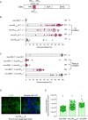
Fig 4. A nuclear localization signal in CifA is necessary for CI, rescue, and protamine levels.
(A) Schematic representation of CifA annotation shows the annotated bNLS with engineered amino acid substitutions and deletions. (B, C) Hatch rate assays assessed both CI (B) and rescue (C) in flies expressing wild-type, transgenic, and mutant cifA. Each dot represents the percent of embryos that hatched from a single male and female pair. Sample size is listed in parentheses. Horizontal bars represent the median. Letters to the right indicate significant differences determined by a Kruskal–Wallis test and Dunn multiple comparison tests. All the p-values are reported in S2 Table. (D) Antibody labeling (green) and DAPI staining of onion stage spermatids in the testes of the bNLS mutant line (cifAΔbNLS) reveals that the deletion ablates CifA’s localization to the nucleus, and CifA thus remains in the surrounding cytoplasm. The imaging experiment was conducted in parallel to nos;cifAB line shown in Fig 1. (E) Mature sperms isolated from seminal vesicles (n = 15) of <8-hour-old males of transgenic cifAΔbnlsB line shows reduced fluorescence indicative of less Protamine deficiency compared to cifAB. To control for any background confounding effects of nos-Gal4VP16 driver line, wMel− fathers were prior crossed to nos- mothers to generate males with nos;wMel− genotype. CMA3 fluorescence levels of sperms isolated from nos;wMel− males were similar to wMel− wild-type lines used in previous assays in this study. Vertical bars represent mean, and error bars represent standard deviation. Letters indicate statistically significant (p < 0.05) differences as determined by multiple comparisons based on a Kruskal–Wallis test and Dunn multiple test correction. All of the p-values are reported in S1 Table, and raw data underlying this figure can be found in S1 Data file. bNLS, bipartite nuclear localization signal; CI, cytoplasmic incompatibility.
Fig 5
Fig 5. CifA, CifB, and the protamine deficiency are transferred with the mature sperm to the female reproductive tract.
(A) Schematic representation of Drosophila melanogaster female reproductive system. Mature oocytes leave the OV and reach the UT, where they can be fertilized prior to being laid. Sperms from males are stored in specialized organs—SP and SR shown in the box, which open into the UT for fertilization to occur. Schematic is created with BioRender.com. (B) Transgenic cifAB-expressing and wMel− males were crossed to wMel− females. Four hours postfertilization, sperms isolated from females were decondensed and immunostained for localizing CifA (green) and CifB (red). DAPI stain (blue) was used to label nuclei. CifA is absent in sperm heads (empty arrowheads) and puctae are seen along the sperm tails (arrows). CifB is present in apical acrosomal tip of all of the sperm heads (solid arrowheads), with more distant signal in the more decondensed sperm nuclei. No Cifs are present in the sperms transferred from wMel− negative control males. (C) Individual sperm intensity quantification shows that protamine deficiency of sperms from wMel+ and transgenic cifAB males persists after transfer in the females compared to wMel− males. Sperm protamine deficiency from transgenic cifAB males also persists in the reproductive tract of wMel+ females. Vertical bars represent mean, and error bars represent standard deviation. Letters indicate statistically significant (p < 0.05) differences as determined by multiple comparisons based on a Kruskal–Wallis test and Dunn multiple test correction. All of the p-values are reported in S1 Table, and raw data underlying this panel can be found in S1 Data file. (D) Representative images of CMA3-stained mature sperms (arrows) transferred from wMel−, wMel+ and transgenic cifAB males in wMel− and wMel+ female reproductive systems are shown. CMA3, chromomycin A3; OV, ovary; SP, spermathecae; SR, seminal receptacle; UT, uterus.
Fig 6
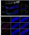
Fig 6. CifA is present in early oogenesis and absent in late-stage egg chambers.
Both CifA and CifB are absent in CI and rescue embryos. (A) Schematic representation of Drosophila melanogaster ovariole at the top illustrates the stages of oogenesis from left to right. Image was created with BioRender.com. Immunostaining assay indicates localization of CifA (green) to the cyst DNA (blue labeled with DAPI) in region 1 of the germarium of Wolbachia-uninfected transgenic cifA line. In wMel+ line, CifA colocalizes with Wolbachia (red) in the germarium, nurse cells and oocyte cytoplasm along 2–8 stages of egg chambers. CifA is absent in stage 10 egg chamber, whereas Wolbachia signals persist. In the transgenic cifA line, we note the observed autofluorescence in green channel outlining the tissue morphology does not signify CifA signals. Ovariole images were manually adjusted in Affinity designer software to align egg chamber stages in the same plane. (B) Immunofluorescence of CifA (green) and CifB (red) in approximately 30- to 60-minute-old embryos obtained from rescue (cifAB male × wMel+ female) and CI crosses (nos;cifAB male × wMel− female). Histone antibody labeling core-histones (magenta) was used as a positive control. Histone signals were detected colocalizing with host DNA, labeled with DAPI (blue), whereas no CifA and CifB signals were detected. Dotted white embryonic periphery is drawn around the embryo shape. White arrows indicate dividing nuclei post fertilization and arrowheads indicate polar bodies. CI, cytoplasmic incompatibility.

Comment in

Similar articles

Cited by

References

    1. Bordenstein SR, Ohara FP, Werren JH. Wolbachia-induced incompatibility precedes other hybrid incompatibilities in Nasonia. Nature. 2001;409:707–10. doi: 10.1038/35055543 - DOI - PubMed
    1. Charlat S, Hurst GDD, Merçot H. Evolutionary consequences of Wolbachia infections. Trends Genet. 2003;19:217–23. doi: 10.1016/S0168-9525(03)00024-6 - DOI - PubMed
    1. Jaenike J, Dyer KA, Cornish C, Minhas MS. Asymmetrical reinforcement and Wolbachia infection in Drosophila. PLoS Biol. 2006;4:1852–62. doi: 10.1371/journal.pbio.0040325 - DOI - PMC - PubMed
    1. Bian G, Xu Y, Lu P, Xie Y, Xi Z. The Endosymbiotic Bacterium Wolbachia Induces Resistance to Dengue Virus in Aedes aegypti. PLoS Pathog. 2010;6:e1000833. doi: 10.1371/journal.ppat.1000833 - DOI - PMC - PubMed
    1. Frentiu FD, Zakir T, Walker T, Popovici J, Pyke AT, van den Hurk A, et al.. Limited Dengue Virus Replication in Field-Collected Aedes aegypti Mosquitoes Infected with Wolbachia. PLoS Negl Trop Dis. 2014;8. doi: 10.1371/journal.pntd.0002688 - DOI - PMC - PubMed

Publication types

LinkOut - more resources