The Promise of Magnetic Resonance Imaging in Radiation Oncology Practice in the Management of Brain, Prostate, and GI Malignancies
- PMID: 35609219
- PMCID: PMC9173575
- DOI: 10.1200/GO.21.00366
The Promise of Magnetic Resonance Imaging in Radiation Oncology Practice in the Management of Brain, Prostate, and GI Malignancies
Abstract
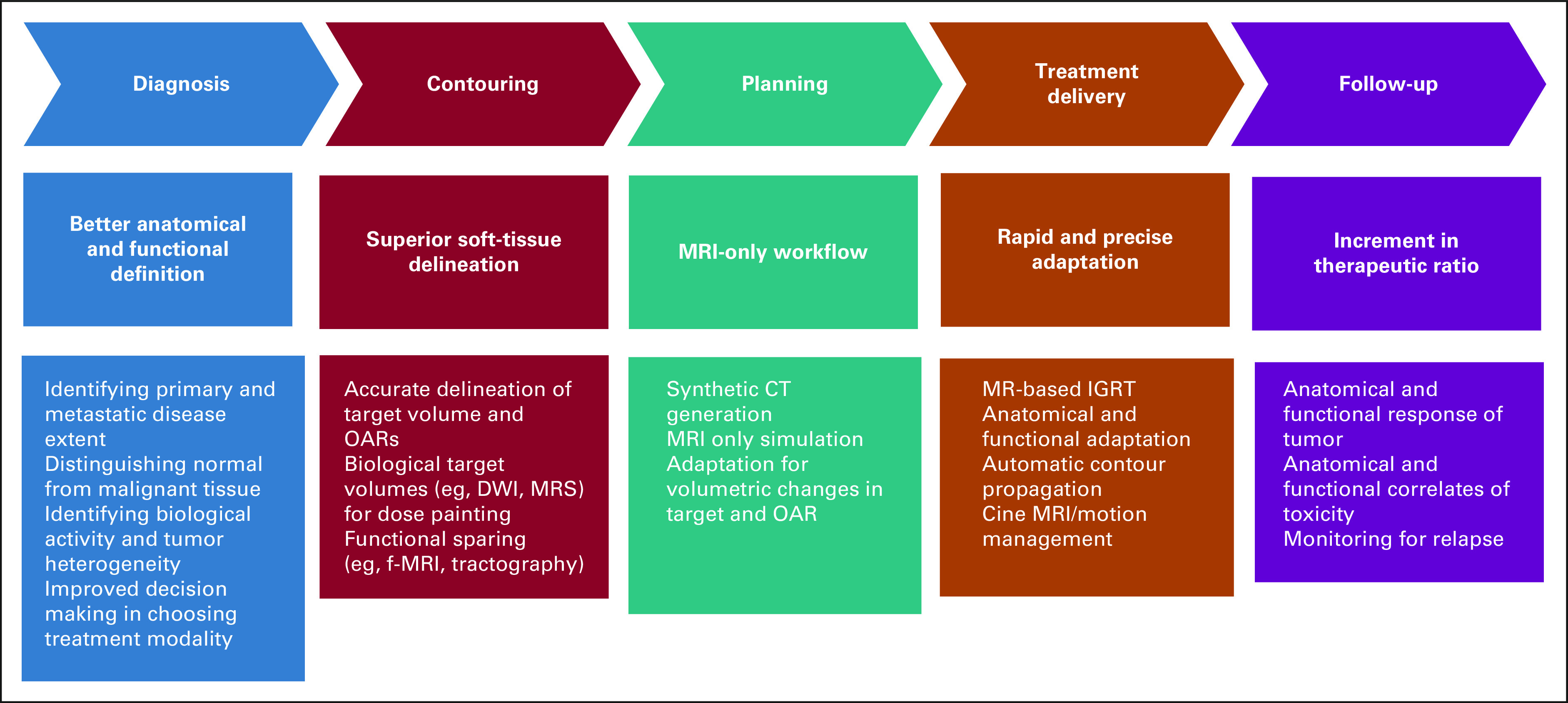
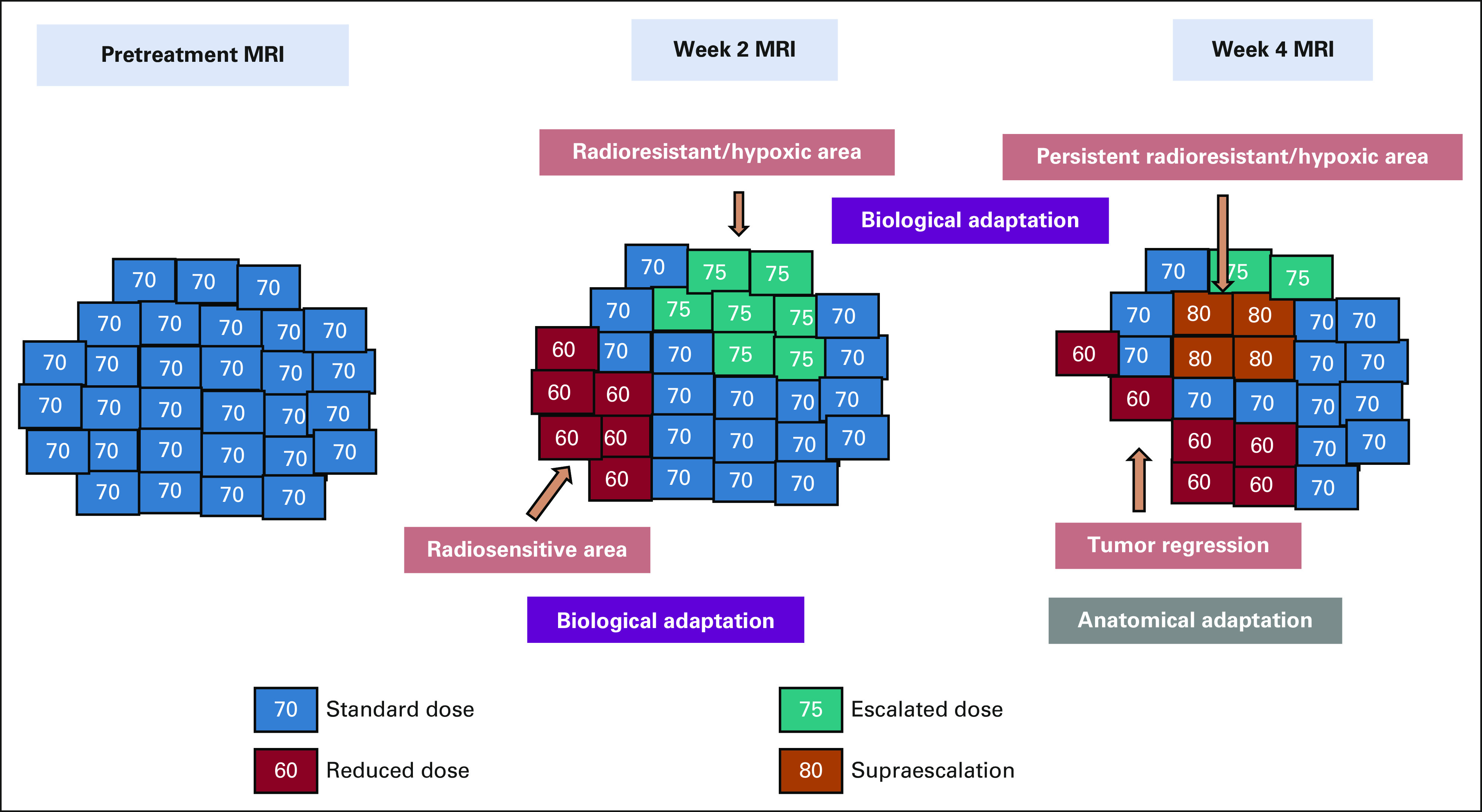
Magnetic resonance imaging (MRI) has a key role to play at multiple steps of the radiotherapy (RT) treatment planning and delivery process. Development of high-precision RT techniques such as intensity-modulated RT, stereotactic ablative RT, and particle beam therapy has enabled oncologists to escalate RT dose to the target while restricting doses to organs at risk (OAR). MRI plays a critical role in target volume delineation in various disease sites, thus ensuring that these high-precision techniques can be safely implemented. Accurate identification of gross disease has also enabled selective dose escalation as a means to widen the therapeutic index. Morphological and functional MRI sequences have also facilitated an understanding of temporal changes in target volumes and OAR during a course of RT, allowing for midtreatment volumetric and biological adaptation. The latest advancement in linear accelerator technology has led to the incorporation of an MRI scanner in the treatment unit. MRI-guided RT provides the opportunity for MRI-only workflow along with online adaptation for either target or OAR or both. MRI plays a key role in post-treatment response evaluation and is an important tool for guiding decision making. In this review, we briefly discuss the RT-related applications of MRI in the management of brain, prostate, and GI malignancies.
Conflict of interest statement
Figures
References
-
- Pooley RA. AAPM/RSNA physics tutorial for residents: Fundamental physics of MR imaging. Radiographics. 2005;25:1087–1099. - PubMed
-
- Edelman RR. The history of MR imaging as seen through the pages of radiology. Radiology. 2014;273(2 suppl):S181–S200. - PubMed
-
- Plewes DB, Kucharczyk W. Physics of MRI: A primer. J Magn Reson Imaging. 2012;35:1038–1054. - PubMed
-
- FitzGerald TJ, Rosen MA, Bishop-Jodoin M. The influence of imaging in the modern practice of radiation oncology. Int J Radiat Oncol Biol Phys. 2018;102:680–682. - PubMed
Publication types
MeSH terms
LinkOut - more resources
Full Text Sources
Medical