. 2023 Apr 1;147(4):390-402.
doi: 10.5858/arpa.2021-0539-RA.

Uncommon Benign Neoplasms and Pseudotumors of the Liver

Affiliations

Uncommon Benign Neoplasms and Pseudotumors of the Liver

Naziheh Assarzadegan et al. Arch Pathol Lab Med. .

Abstract

Context.—: The most common benign hepatic mass-forming lesions often display fairly specific imaging characteristics, whereas less familiar, rarer benign neoplasms and pseudotumors may pose a diagnostic challenge in clinical, radiology, and pathology practice because of either their rarity or their unusual features.

Objective.—: To review a selection of pseudotumors and unusual benign hepatic neoplasms encountered in consultation practices with a focus on nonepithelial tumors.

Data sources.—: Sources include English-language literature and personal experiences.

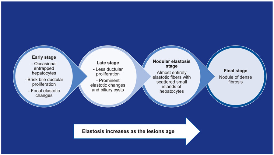
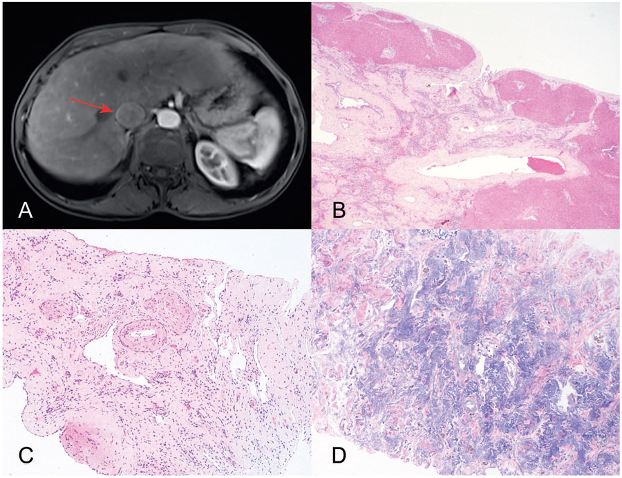
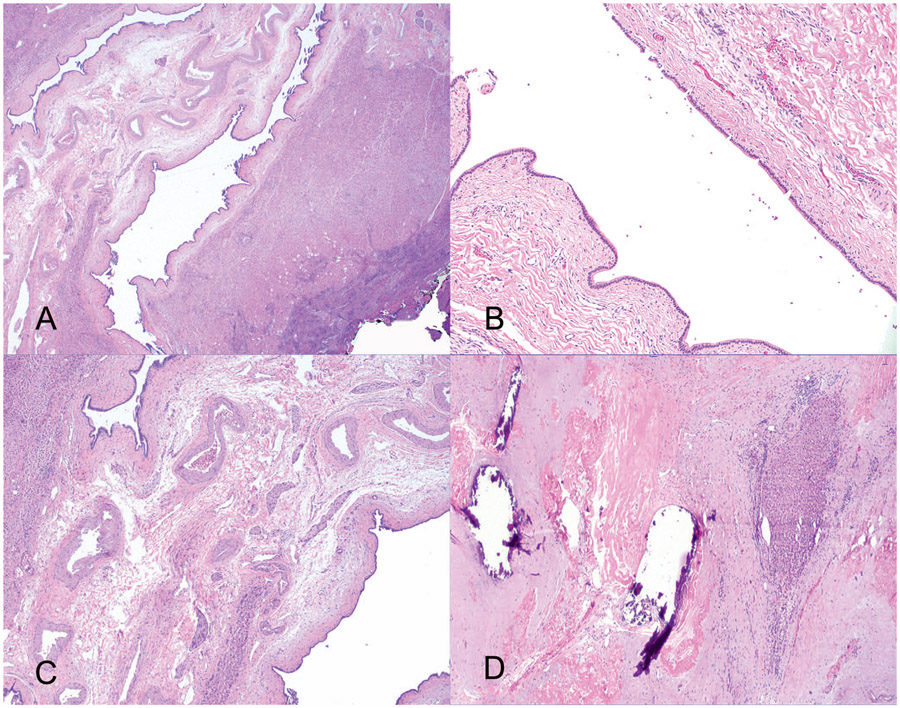
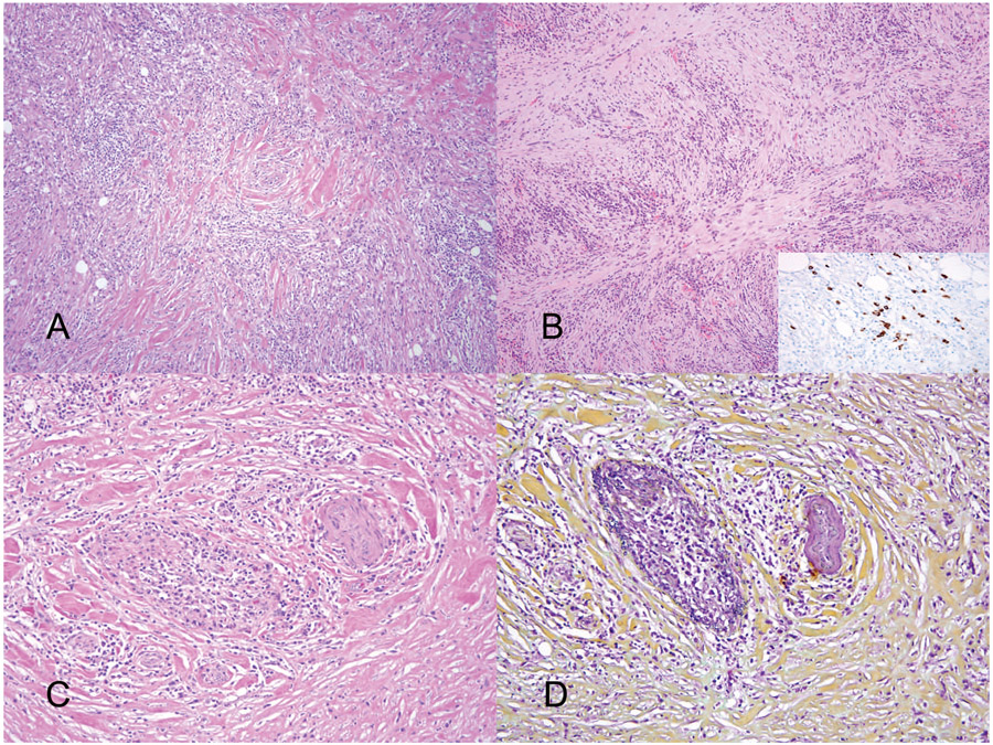
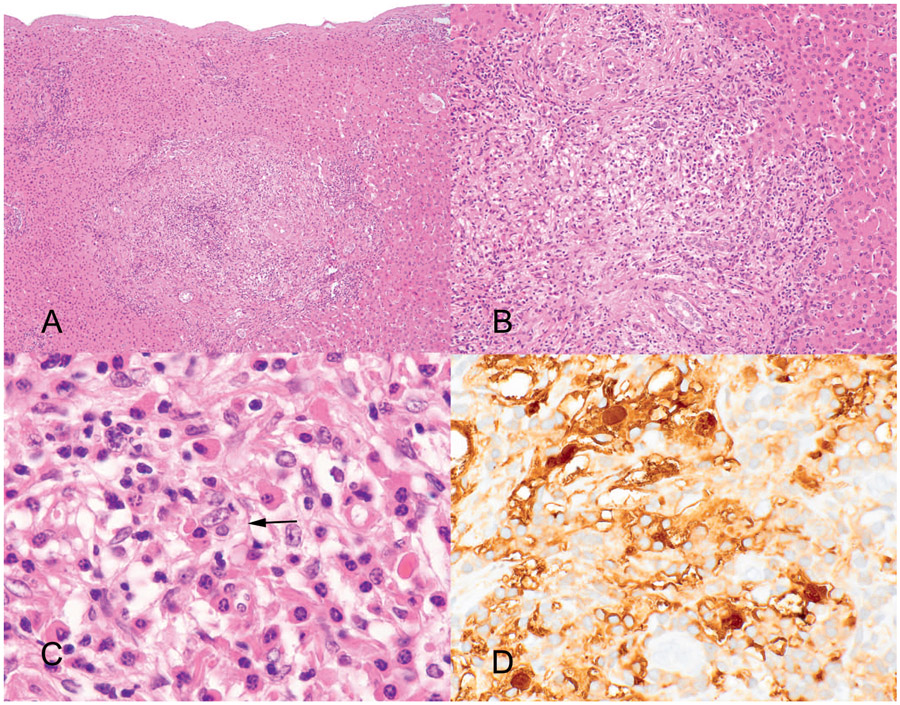
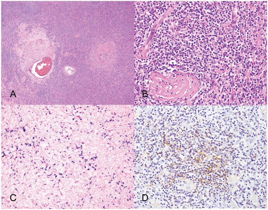
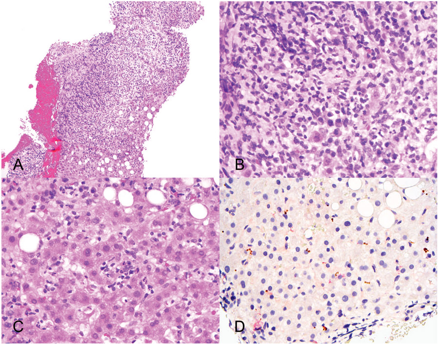
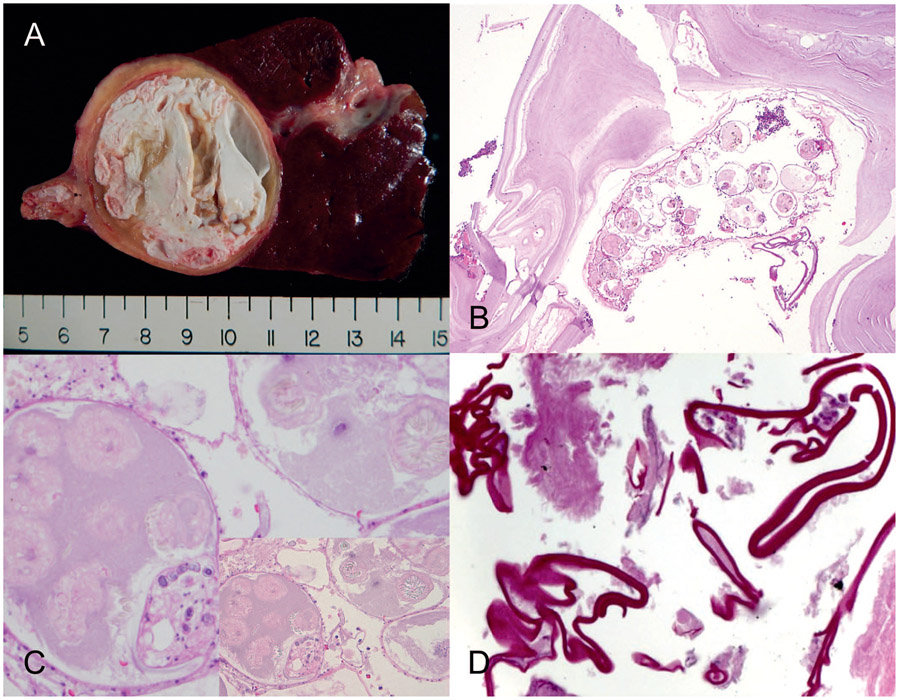
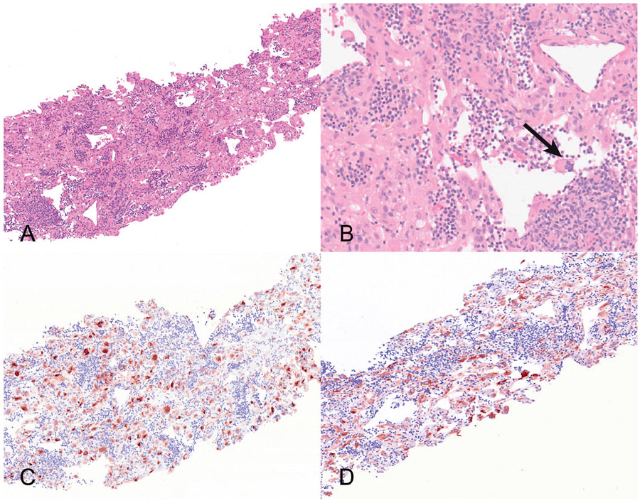
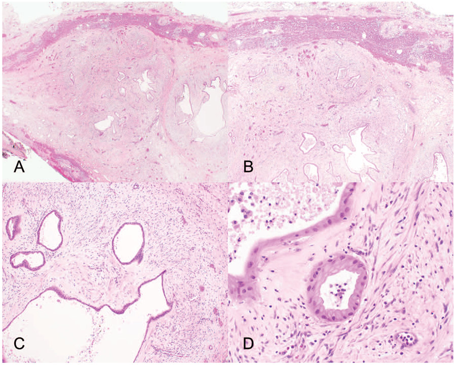
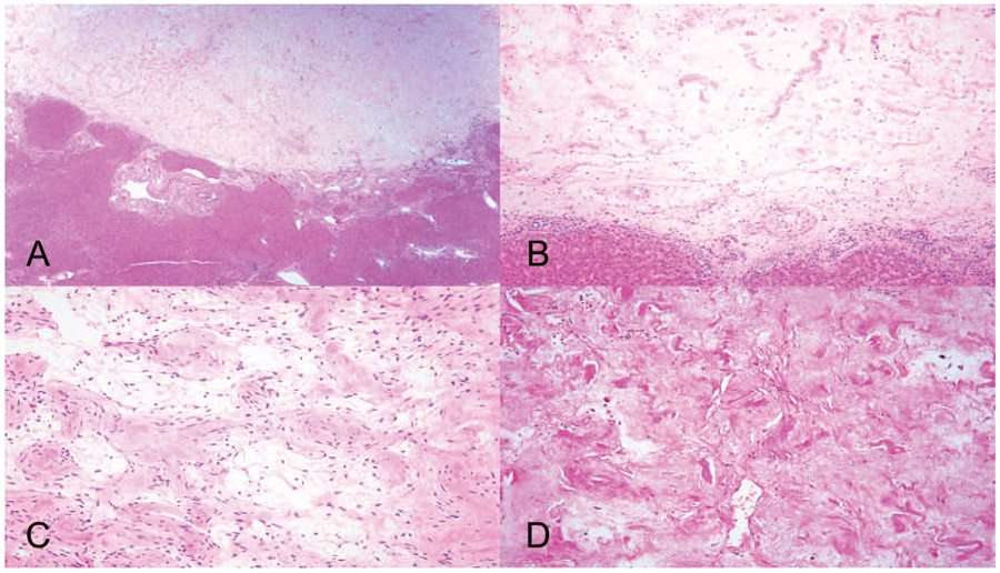
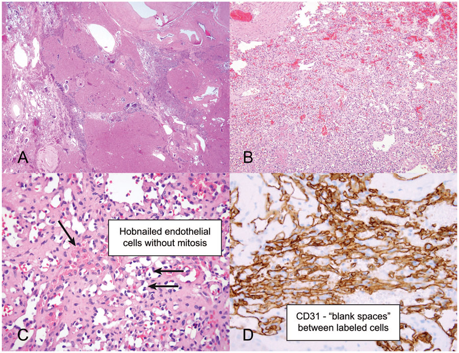
Conclusions.—: Several benign conditions (namely, segmental atrophy, infections, immunoglobulin G4 [IgG4]-related sclerosing disease, angiomyolipoma, mesenchymal hamartoma, and various vascular lesions) can lead to formation of hepatic masses. Because of their rarity and underrecognition, such lesions are often diagnostically challenging. Awareness of hepatic pseudotumors and various rare hepatic neoplasms and their potential mimics can forestall misdiagnosis and inappropriate management.


Conflict of interest statement

The authors have no relevant financial interest in the products or companies described in this article.

Figures

Figure 1.
Figure 1.
Segmental atrophy stages.
Figure 2.
Figure 2.
Segmental atrophy (SA). A, Magnetic resonance image shows an abnormal appearance of the inferior right hepatic lobe/portions of segment 6 near the gallbladder fossa (arrow), with a subtle area of focal capsular retraction concerning for cholangiocarcinoma. B, Histologic examination shows a subcapsular lesion showing residual entrapped hepatocytes, inflammation, and bile ductular proliferation. C, SA at its later stages shows increased elastosis as well as thick-walled blood vessels. D, Elastin stain highlights the elastic fibers (hematoxylin-eosin, original magnifications ×2 [B] and ×10 [C]; elastin, original magnification ×400 [D]).
Figure 3.
Figure 3.
Segmental atrophy (SA). This is an 11-cm subcapsular lesion clinically concerning for mucinous cystic neoplasm or intraductal papillary neoplasm of bile ducts. A and B, At low magnification, a retention-type biliary cyst, lined with bland biliary-type epithelium without ovarian-type stroma, dominates the histology at least focally suggesting other types of cystic biliary tract disease. C and D, Adjacent to the cyst and in other areas of the lesion, classic features of SA with thick-walled blood vessels and elastosis are seen (hematoxylin-eosin, original magnifications ×2 [A], ×10 [B], ×100 [C], and ×200 [D]).
Figure 4.
Figure 4.
Immunoglobulin G4 (IgG4)–related sclerosing disease. A, Low magnification shows a fibroinflammatory lesion with areas of storiform fibrosis. B, Numerous plasma cells express IgG4 on immunostain (inset). C and D, Obliterative phlebitis is characterized by a damaged vein adjacent to an artery, as highlighted on (D) Movat stain (hematoxylin-eosin, original magnifications ×2 [A] and ×10 [B and C]; IgG4 immunostain, original magnification ×400 [B, inset]; original magnification ×400 [D]).
Figure 5.
Figure 5.
Rosai-Dorfman disease (RDD) involving the liver. A, Low magnification shows a fibroinflammatory process predominantly centered within the portal tracts. B, Higher magnification shows a mixed inflammatory infiltrate rich in histiocytes and plasma cells. C, Emperipolesis is indicated by the arrow. D, Strong S100 protein reactivity with prominent emperipolesis. The ingested lymphocytes appear pale within the stained cytoplasm of the lesional histiocytes. The nuclei of the RDD cells express S100 protein (hematoxylin-eosin, original magnifications ×10 [A], ×100 [B], and ×200 [C]; S100 immunostain, original magnification ×400 [D]).
Figure 6.
Figure 6.
Inflammatory pseudotumor-like follicular/fibroblastic dendritic cell sarcoma. A and B, The lesion is composed of spindle cells heavily admixed with lymphocytes, histiocytes, and plasma cells. Note the fibrinoid deposits in the wall of blood vessels. C, Immunostaining for CD21 shows focal, patchy staining. D, The spindle cells showed strong nuclear in situ labeling for Epstein-Barr virus–encoded early nuclear RNAs (EBER), whereas the background inflammatory cells are uniformly negative (hematoxylin-eosin, original magnifications ×10 [A] and ×200 [B]; CD21 immunostain, original magnification ×200 [C]; EBER in situ hybridization, original magnification ×200 [D]).
Figure 7.
Figure 7.
Syphilis presenting as a liver pseudotumor. A, Low magnification shows a fibroinflammatory process. B, Higher magnification shows the abundance of plasma cells in the inflammatory infiltrate. C, Scattered mild lobular lymphocytic and neutrophilic inflammation in the background liver. D, Treponema pallidum immunostain reveals the presence of spiral organisms (hematoxylin-eosin, original magnifications ×2 [A] and ×10 [B and C]; original magnification ×400 [D]). Images courtesy of Lysandra Voltaggio, MD.
Figure 8.
Figure 8.
Echinococcosis granulosus. A, The wedge resection shows a unilocular cystic lesion with a fibrous rim filled with grungy white material. B, Histologic sections showed predominantly acellular hyalinized material and remnants of degenerated parasites with smaller daughter cysts. C, Higher magnification shows refractile hooklets, which are best seen with the condenser down and with refracted light (inset). D, Periodic acid–Schiff stain highlighting the cyst wall (hematoxylin-eosin, original magnifications ×10 [B] and ×200 [C]; original magnification ×200 [D]). Images courtesy of Michael Feely, DO.
Figure 9.
Figure 9.
Fat-poor angiomyolipoma. A, Low magnification shows a richly vascular lesion composed of epithelioid cells with abundant eosinophilic cytoplasm. B, Extramedullary hematopoiesis (arrow). C and D, Immunostains for HMB-45 and Melan-A (hematoxylin-eosin, original magnifications ×10 [A] and ×100 [B]; HMB-45 and Melan-A, original magnification ×200 [C and D]). Images courtesy of Richard Kirsch, MD.
Figure 10.
Figure 10.
Mesenchymal hamartoma. A and B, Low magnifications show a biphasic lesion composed of numerous bile ducts in a loose mesenchymal stroma with interspersed islands of normal hepatocytes. C, Higher magnification shows both epithelial and mesenchymal components. D, Both components have bland nuclei with no cytologic atypia or mitoses (hematoxylin-eosin, original magnifications ×10 [A and B], ×100 [C], and ×200 [D]).
Figure 11.
Figure 11.
Sclerosed hemangioma. A, Hemangioma with predominant sclerosis and near-complete obliteration of the vascular spaces. B, Higher magnification of remnant vessels. C, Ghostly remnants of vascular spaces. D, Negative elastin stain (hematoxylin-eosin, original magnifications ×10 [A], ×100 [B], and ×200 [C]; original magnification ×400 [D]).
Figure 12.
Figure 12.
Anastomosing hemangioma. A, Infiltrative vascular lesion at low magnification. B, Medium power shows an anastomosing vascular lesion. C, Higher magnification shows scattered hyaline globules and hobnailed endothelial cells without mitosis. D, CD31 immunostain highlighting the anastomosing pattern and blank spaces between the labeled cells (hematoxylin-eosin, original magnifications ×2 [A], ×10 [B], and ×100 [C]; original magnification ×400 [D]).

References

    1. Singhi AD, Maklouf HR, Mehrotra AK, et al. Segmental atrophy of the liver: a distinctive pseudotumor of the liver with variable histologic appearances. Am J Surg Pathol. 2011;35(3):364–371. doi: 10.1097/PAS.0b013e31820b0603 - DOI - PubMed
    1. Bedada AG, Sreekumaran MI, Azzie G. Segmental atrophy of the liver in a child: case report and review of the literature. S Afr J Child Health. 2019;13(2): 100–101. doi:10.7196/SAJCH.2019.v13i2.1574 - DOI
    1. Assarzadegan N, Estevez-Castro R, Hutchings D, et al. Segmental atrophy (pseudotumor) of the liver: a marker of cardiovascular disease that mimics neoplasia. Mod Pathol. 2020;33(suppl 2):1500.
    1. Torbenson M, Yasir S, Anders R, et al. Regenerative hepatic pseudotumor: a new pseudotumor of the liver. Hum Pathol. 2020;99:43–52. doi:10.1016/j.humpath.2020.03.010 - DOI - PMC - PubMed
    1. Divatia M, Kim SA, Ro JY. IgG4-related sclerosing disease, an emerging entity: a review of a multi-system disease. Yonsei Med J. 2012;53(1):15–34. doi: 10.3349/ymj.2012.53.1.15 - DOI - PMC - PubMed

MeSH terms