. 2022 Jul:53:102338.
doi: 10.1016/j.redox.2022.102338. Epub 2022 May 16.

Mitigation of doxorubicin-induced cardiotoxicity with an H2O2-Activated, H2S-Donating hybrid prodrug

Affiliations

Mitigation of doxorubicin-induced cardiotoxicity with an H2O2-Activated, H2S-Donating hybrid prodrug

Qiwei Hu et al. Redox Biol. 2022 Jul.

Abstract

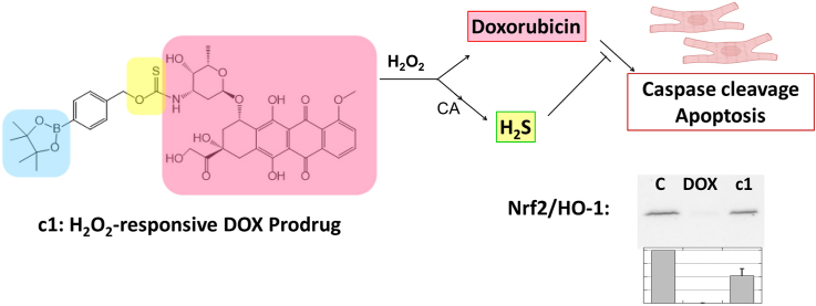
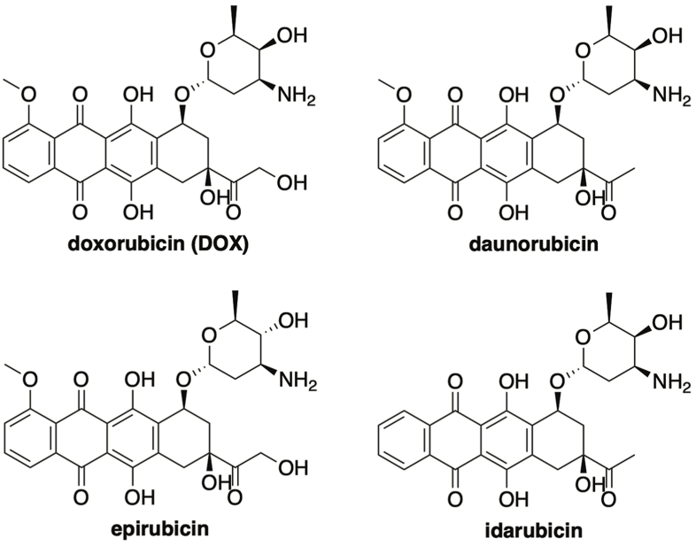
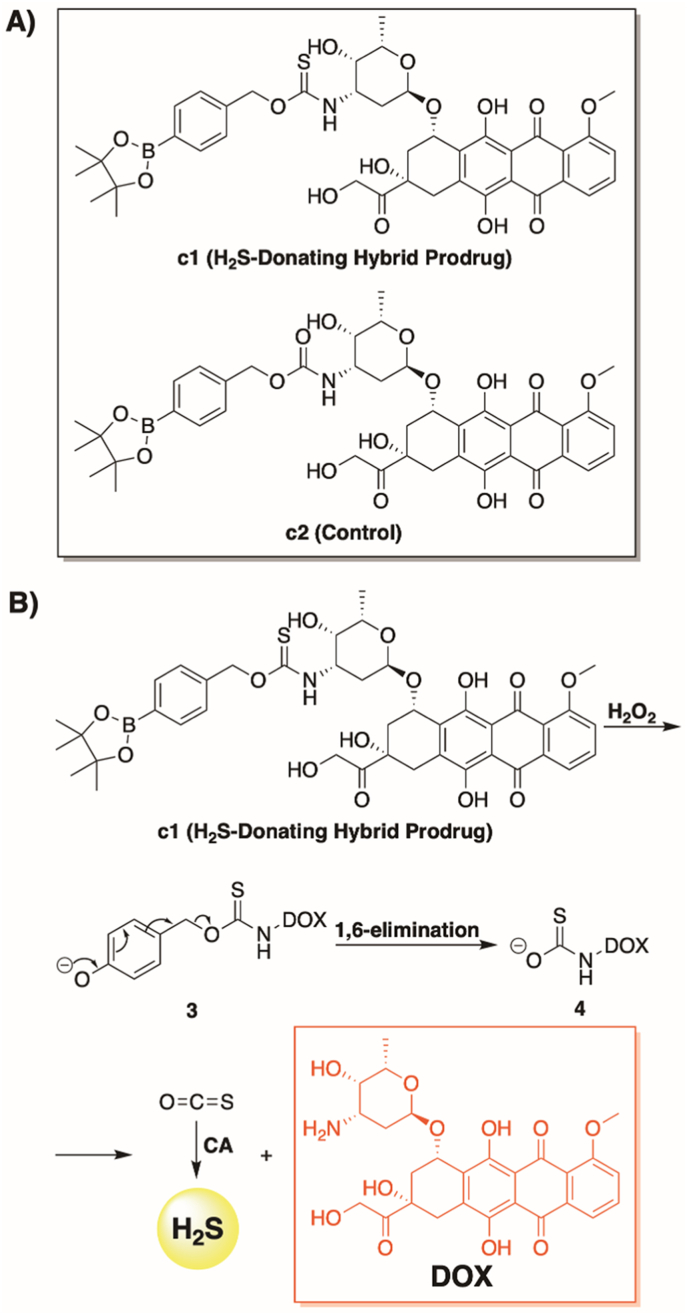
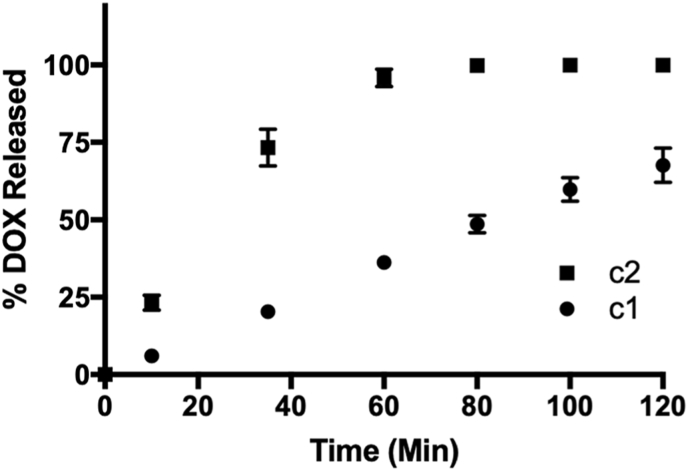
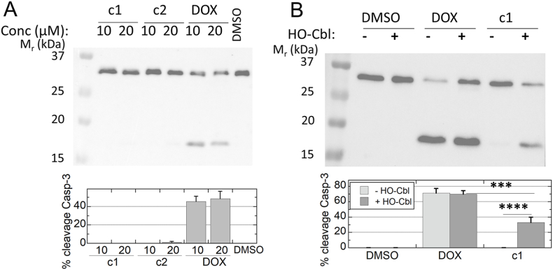
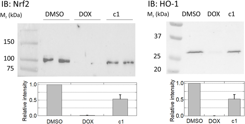
Doxorubicin (DOX) is one of the most effective anticancer agents in clinical oncology. Its continued use, however, is severely limited by its dose-dependent cardiotoxicity which stems, in part, from its overproduction of reactive oxygen species (ROS) and often manifests itself as full-blown cardiomyopathy in patients, years after the cessation of treatment. Therefore, identifying DOX analogs, or prodrugs, with a diminished cardiotoxic profile is highly desirable. Herein, we describe a novel, H2O2-responsive DOX hybrid codrug (mutual prodrug) that has been rationally designed to concurrently liberate hydrogen sulfide (H2S), a purported cardioprotectant with anticancer activity, in an effort to maintain the antitumor effects of DOX while simultaneously reducing its cardiotoxic side effects. Experiments with cardiomyoblast cells in culture demonstrated a rapid accumulation of prodrug into the cells, but diminished apoptotic effects compared with DOX, dependent upon its release of H2S. Cells treated with the prodrug exhibited significantly higher Nrf2 activation relative to DOX-treated cells. Preliminary indications, using a mouse triple-negative breast cancer cell line sensitive to DOX treatment, are that the prodrug maintains considerable toxicity against the tumor-inducing cell line, suggesting significant promise for this prodrug as a cardioprotective chemotherapeutic to replace DOX.

Keywords: Cardiotoxicity; Chemotherapeutic; Doxorubicin; Hydrogen peroxide; Hydrogen sulfide.


Conflict of interest statement

The authors declare the following financial interests/personal relationships which may be considered as potential competing interests: John C. Lukesh reports financial support was provided by National Science Foundation. Leslie B. Poole reports financial support was provided by National Institute of General Medical Sciences. John C. Lukesh and Leslie B. Poole have a provisional patent pending to Wake Forest University.

Figures

Image 1
Graphical abstract
Fig. 1
Fig. 1
Anthracycline antibiotics.
Fig. 2
Fig. 2
Prodrug structures and H2O2-dependent release pathway, (A) Prodrugs assessed in this study. (B) Proposed mechanism for the simultaneous release of H2S and DOX from c1 in response to H2O2. CA = carbonic anhydrase.
Fig. 3
Fig. 3
Time-course for DOX release from prodrugs (10 μM) in ammonium bicarbonate buffer (0.1 M, pH 7.4) at room temperature and in the presence of H2O2 (10 μM). A calibration curve was used to determine the concentration of free DOX at each time point by LCMS. Plotted as the mean ± standard error of the mean (SEM) from three independent experiments.
Fig. 4
Fig. 4
Percentage of released DOX from c1 in response to various biological analytes during an 80 min incubation period at room temperature: (1) ammonium bicarbonate buffer (0.1 M, pH 7.5), (2) 10 μM H2O2, (3) 100 μM cysteine, (4) 100 μM homocysteine, (5) 1 mM glutathione, (6) 10 μM glutathione disulfide, (7) 10 μM sodium nitrite, (8) 10 μM sodium hypochlorite, (9) 10 μM superoxide, (10) 10 μM peroxynitrite. A calibration curve was used to determine the concentration of free DOX in response to each analyte. Plotted as the mean ± SEM from three independent experiments.
Fig. 5
Fig. 5
Methylene blue assay depicting the time-dependent release of H2S from c1 (40 μM) while in the presence of H2O2 (40 μM) and carbonic anhydrase (CA). Plotted as the mean ± SEM from three independent experiments. Data were collected in the presence (circles) or absence (squares) of H2O2.
Fig. 6
Fig. 6
Uptake of DOX and prodrugs c1 and c2 by H9C2 cardiomyoblasts. H9C2 cells cultures grown overnight on chambered coverslips in media with 10% serum were switched to Fluorobrite DMEM imaging media supplemented with 5% serum and prepared for live-cell imaging on a Zeiss LSM 880 confocal microscope, then 10 μM of c1, c2, or DOX was added and images were taken every 10 min (averaged every 30 min) for 18 h. Approximately 20 cells present in each field of view were averaged for each sample and time point; normalized and averaged data from two replicates each ± SEM were included for c1 and DOX. An earlier independent trial yielded very similar results (Fig. S4).
Fig. 7
Fig. 7
Cytotoxicity of DOX and prodrugs in H9C2 cardiomyoblasts. Media from cells exposed for 24 or 48 h to 10 or 20 μM (A and B, respectively) of c1, c2, DOX or vehicle (DMSO, final concentration 0.1% in all samples) was centrifuged to remove cells and cell debris, and supernatants were assessed spectrophotometrically by lactose dehydrogenase (LDH) assay to evaluate release into the media as a measure of cytotoxicity (n = 6 or more). *, p < 0.05; **, p < 0.01; ***, p < 0.001; ****, p < 0.0001.
Fig. 8
Fig. 8
Caspase cleavage monitored by an antibody against total caspase-3 demonstrates the H2S -dependent protection exhibited by c1 against DOX-mediated apoptotic signaling. H9C2 cells in culture were treated for 24 h with c1, c2, DOX or vehicle (DMSO), then harvested into lysis buffer and immunoblotted for caspase-3. Data analyzed by ImageJ were used to assess the percent of the two bands present as the lower band. A. At 24 h, only DOX treatment causes caspase-3 cleavage (n = 3). B. When 100 mM hydroxocobalamin is added 5 min prior to 10 μM drug treatments, c1-treated cells exhibit much more cleavage of caspase-3 than in its absence, whereas cleavage due to DOX treatment is unchanged (n = 7); ***, p < 0.001; ****, p < 0.0001.
Fig. 9
Fig. 9
c1-treated cardiomyoblasts do not shut down Nrf2 activation as DOX does. Immunoblots for the transcriptional regulator Nrf2 (left) and one of its downstream targets, HO-1 (right), demonstrate stabilization of Nrf2 with concomitant expression of HO-1 in DMSO-treated samples; DOX treatment completely suppressed both, while c1 treatment was only moderately suppressive (n = 4 and n = 3 for Nrf2 and HO-1, respectively). The bar graphs below represent the mean ± SEM. Results were statistically different between c1 and DOX in both cases (p = 0.002 and p = 0.019), and more marginally so between c1 and DMSO (p = 0.011 and p. = 0.025), for Nrf2 and HO-1, respectively.

References

    1. Minotti G., Menna P., Salvatorelli E., Cairo G., Gianni L. Anthracyclines: molecular advances and pharmacologic developments in antitumor activity and cardiotoxicity. Pharmacol. Rev. 2004;56(2):185–229. doi: 10.1124/pr.56.2.6. - DOI - PubMed
    1. Arcamone F., Cassinelli G., Fantini G., Grein A., Orezzi P., Pol C., Spalla C. Adriamycin, 14-hydroxydaimomycin, a new antitumor antibiotic FromS. Peucetius Var.Caesius. Biotechnol. Bioeng. 1969;11(6):1101–1110. doi: 10.1002/bit.260110607. - DOI - PubMed
    1. Sun J., Wei Q., Zhou Y., Wang J., Liu Q., Xu H. A systematic analysis of FDA-approved anticancer drugs. BMC Syst. Biol. 2017;11(S5):87. doi: 10.1186/s12918-017-0464-7. - DOI - PMC - PubMed
    1. Sritharan S., Sivalingam N. A comprehensive review on time-tested anticancer drug doxorubicin. Life Sci. 2021;278:119527. doi: 10.1016/j.lfs.2021.119527. - DOI - PubMed
    1. Gewirtz D.A. Critical evaluation of the mechanisms of action proposed for the antitumor effects of the anthracycline antibiotics adriamycin and daunorubicin. Biochem. Pharmacol. 1999;57(7):727–741. doi: 10.1016/S0006-2952(98)00307-4. - DOI - PubMed

Publication types

LinkOut - more resources