

Refolding of lid subdomain of SARS-CoV-2 nsp14 upon nsp10 interaction releases exonuclease activity
- PMID: 35609600
- PMCID: PMC9125827
- DOI: 10.1016/j.str.2022.04.014
Refolding of lid subdomain of SARS-CoV-2 nsp14 upon nsp10 interaction releases exonuclease activity
Abstract
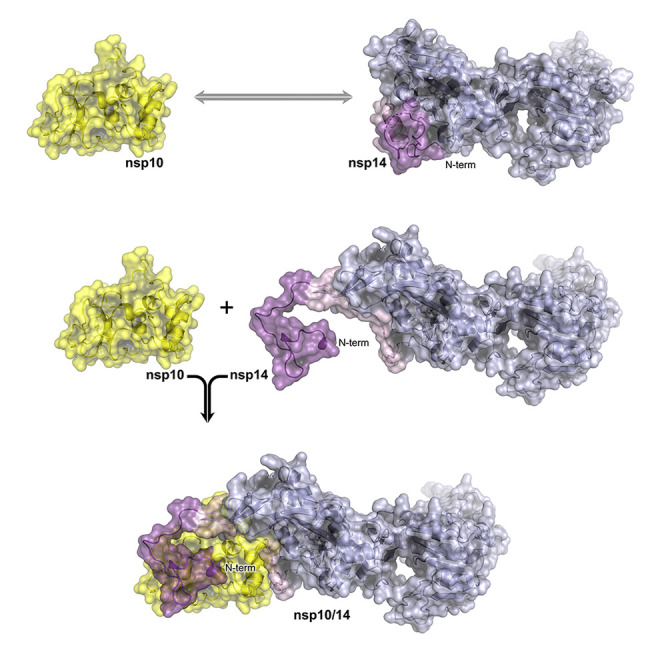
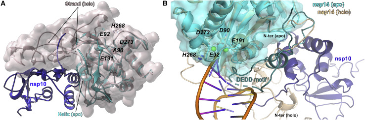
During RNA replication, coronaviruses require proofreading to maintain the integrity of their large genomes. Nsp14 associates with viral polymerase complex to excise the mismatched nucleotides. Aside from the exonuclease activity, nsp14 methyltransferase domain mediates cap methylation, facilitating translation initiation and protecting viral RNA from recognition by the innate immune sensors. The nsp14 exonuclease activity is modulated by a protein co-factor nsp10. While the nsp10/nsp14 complex structure is available, the mechanistic basis for nsp10-mediated modulation remains unclear in the absence of the nsp14 structure. Here, we provide a crystal structure of nsp14 in an apo-form. Comparative analysis of the apo- and nsp10-bound structures explain the modulatory role of the co-factor protein and reveal the allosteric nsp14 control mechanism essential for drug discovery. Further, the flexibility of the N-terminal lid of the severe acute respiratory syndrome coronavirus 2 (SARS-CoV-2) nsp14 structure presented in this study rationalizes the recently proposed idea of nsp14/nsp10/nsp16 ternary complex.
Keywords: SAH; SARS-CoV-2; XRD; coronavirus; methylation; methyltransferase; nsp10; nsp14; proof-reading; structure.
Copyright © 2022 The Author(s). Published by Elsevier Ltd.. All rights reserved.
Conflict of interest statement
Declaration of interests The authors declare no competing interests.
Figures
References
-
- Baddock H.T., Brolih S., Yosaatmadja Y., Ratnaweera M., Bielinski M., Swift L.P., Cruz-Migoni A., Fan H., Keown J.R., Walker A.P., et al. Characterization of the SARS-CoV-2 ExoN (nsp14ExoN-nsp10) complex: implications for its role in viral genome stability and inhibitor identification. Nucleic Acids Res. 2022;50:1484–1500. doi: 10.1093/nar/gkab1303. - DOI - PMC - PubMed
-
- Bouvet M., Imbert I., Subissi L., Gluais L., Canard B., Decroly E. RNA 3'-end mismatch excision by the severe acute respiratory syndrome coronavirus nonstructural protein nsp10/nsp14 exoribonuclease complex. Proc. Natl. Acad. Sci. U S A. 2012;109:9372–9377. doi: 10.1073/pnas.1201130109. - DOI - PMC - PubMed
-
- Devkota K., Schapira M., Perveen S., Khalili Yazdi A., Li F., Chau I., Ghiabi P., Hajian T., Loppnau P., Bolotokova A., et al. Probing the SAM binding site of SARS-CoV-2 Nsp14 in vitro using SAM competitive inhibitors guides developing selective bisubstrate inhibitors. SLAS Discov. 2021;26:1200–1211. doi: 10.1177/24725552211026261. - DOI - PMC - PubMed
Publication types
MeSH terms
Substances
LinkOut - more resources
Full Text Sources
Miscellaneous
