Review
. 2022 Aug 15:257:119329.
doi: 10.1016/j.neuroimage.2022.119329. Epub 2022 May 21.

Imaging perivascular space structure and function using brain MRI

Affiliations
Review

Imaging perivascular space structure and function using brain MRI

Giuseppe Barisano et al. Neuroimage. .

Abstract

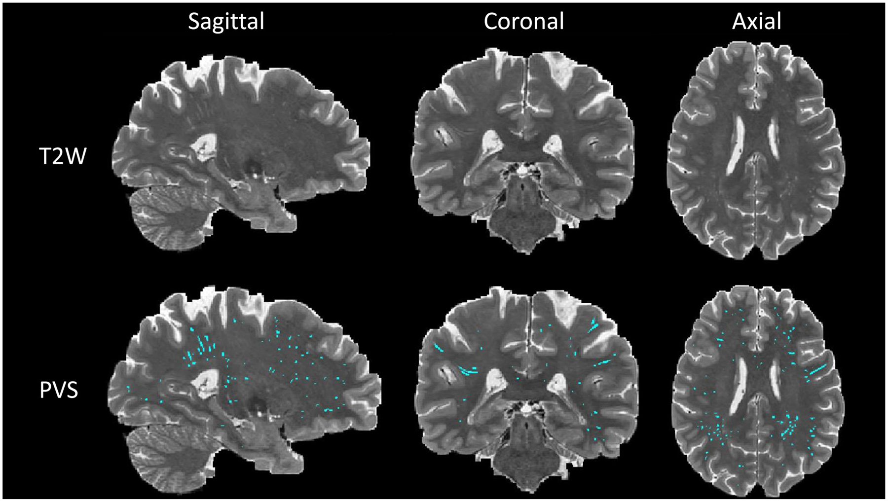
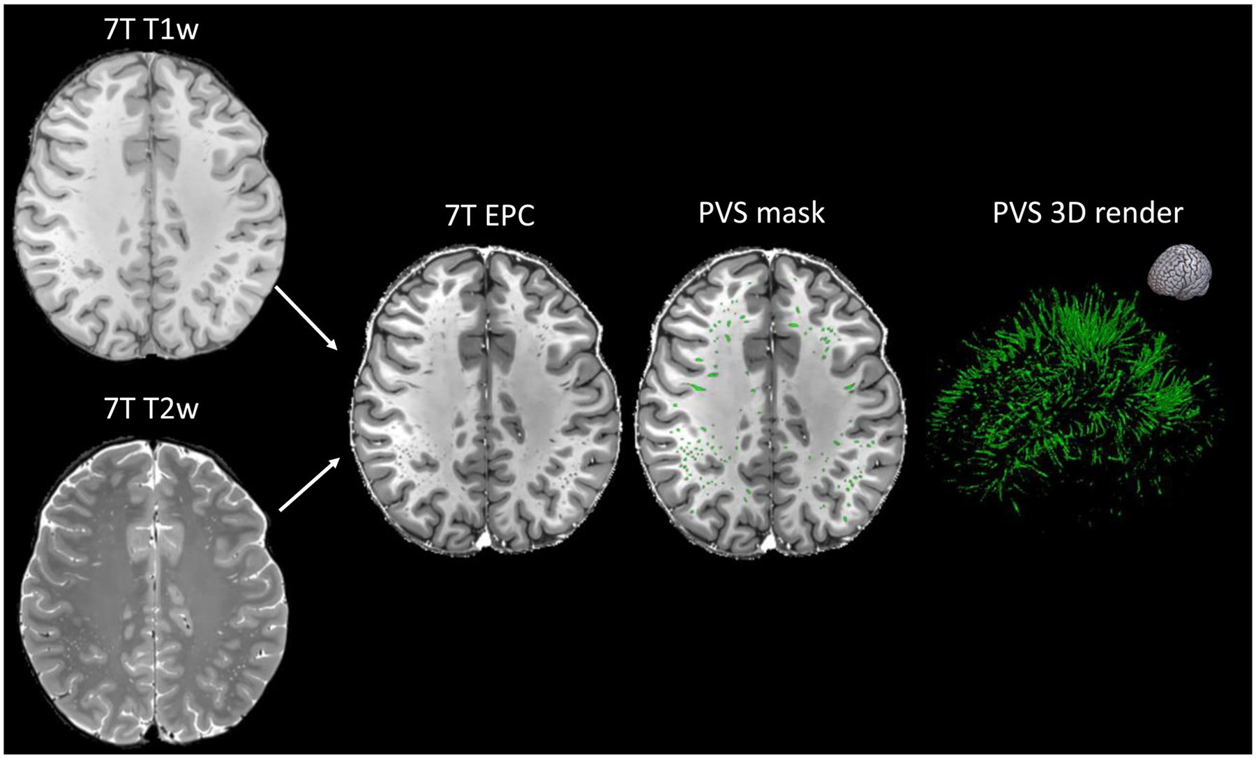
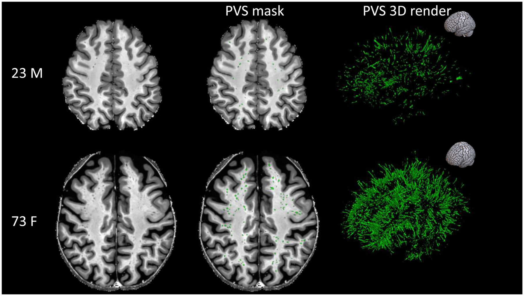
In this article, we provide an overview of current neuroimaging methods for studying perivascular spaces (PVS) in humans using brain MRI. In recent years, an increasing number of studies highlighted the role of PVS in cerebrospinal/interstial fluid circulation and clearance of cerebral waste products and their association with neurological diseases. Novel strategies and techniques have been introduced to improve the quantification of PVS and to investigate their function and morphological features in physiological and pathological conditions. After a brief introduction on the anatomy and physiology of PVS, we examine the latest technological developments to quantitatively analyze the structure and function of PVS in humans with MRI. We describe the applications, advantages, and limitations of these methods, providing guidance and suggestions on the acquisition protocols and analysis techniques that can be applied to study PVS in vivo. Finally, we review the human neuroimaging studies on PVS across the normative lifespan and in the context of neurological disorders.

Keywords: Diffusion MRI; Perivascular spaces; Structural MRI; Ultra-high field MRI.


Conflict of interest statement

Declaration of Competing Interest The authors declare that they have no known competing financial interests or personal relationships that could have appeared to influence the work reported in this paper.

Figures

Fig. 1.
Fig. 1.
Examples of morphological characteristics computed on two MRI-visible PVS with different shapes: curved (PVS1) and straight (PVS2) (top and bottom rows, respectively). (a) MRI scans acquired from a healthy 25-year-old female subject at 7T with the protocol in Table 2. Masks (cyan) of the PVS examples were overlaid on the right images. (b) 3D representation of the PVS voxels (cyan cubes with black dots). (c) 3D representation of the PVS path (red line), i.e., the longest line connecting any two PVS voxels (cubes with black dots), and the voxels of the PVS path (cubes with red asterisks), computed to measure the mean cross-sectional diameter similar to Zong et al. (2016). The PVS voxels overlapping with the voxels of the corresponding PVS path are indicated by the cubes with both a red asterisk and a black dot. The overlap is higher in PVS2 than PVS1. Each PVS voxel is associated to the nearest voxel on the PVS path (cubes with the same color) and they are used to calculate the cross-sectional diameter. The voxels on the PVS path not associated to any PVS voxel are indicated by white cubes with red asterisks, while the last voxels on the PVS path are indicated by white cubes with both red asterisks and black dots. The cross-sectional diameter in each colored voxel on the PVS path (non-white cubes with red asterisks) is computed as per formula 1, where N is the number of PVS voxels associated to that voxel on the PVS path (i.e., PVS voxels with the same color as the corresponding voxel on the PVS path) and 1 is the mean distance between that voxel on the PVS path and its two neighbors voxels on the PVS path. (d) 3D representation of the PVS linearity, calculated similar to Boespflug et al. (2018): the blue line is the best fit line computed with singular value decomposition and the red lines are the norms connecting the center of each PVS voxel to the best fit line. The longer the norms are (i.e., the more the centers of the PVS voxels are distant from the best fit line), the less linear the PVS is. In this example, PVS1 is less linear than PVS2. (e) 3D representation of solidity based on the convex hull (red polygon) and its vertices (red dots on the voxels). The convex hull in PVS1 includes a large region with no PVS voxels, whereas in PVS2 the convex hull includes mostly PVS voxels. Solidity of PVS1 is therefore lower compared with PVS2.
Fig. 2.
Fig. 2.
PVS map segmented by Unet algorithm. First row is the T2-weighted modality of one subject with sagittal, coronal, and axial view. Second row is the PVS map (cyan) overlaid on T2-weighted modality from the same subject. PVS map was generated using deep learning model Unet which was trained by supervised learning manner with manually annotated PVS map.
Fig. 3.
Fig. 3.
Example of enhanced PVS contrast (EPC) post-processing technique (Sepehrband et al., 2019a) applied on T1-weighted and T2-weighted images acquired from a healthy 25-year-old female subject at 7T MRI with the protocol in Table 2. The T1-weighted, T2-weighted, and EPC images are shown on the left column and the PVS mask was overlaid in the center (green). The image on the right is the corresponding 3D map of the PVS masks. The orientation of the 3D map is reported on the top right corner.
Fig. 4.
Fig. 4.
Examples showing the higher number and total volume of MRI-visible perivascular spaces (PVS) in healthy participants with different ages. The participant on the top is a 23-year-old male, while the participant on the bottom is a 73-year-old female. The MRI scans are shown on the left column and the PVS masks were overlaid in the center (green). The images on the right are the corresponding 3D maps of the PVS masks. The orientation of the 3D maps is reported on the top right corner.

References

    1. Absinta M, Ha SK, Nair G, Sati P, Luciano NJ, Palisoc M, Louveau A, Zaghloul KA, Pittaluga S, Kipnis J, Reich DS, 2017. Human and nonhuman primate meninges harbor lymphatic vessels that can be visualized noninvasively by MRI. Elife 6, 1–15. doi: 10.7554/eLife.29738.001. - DOI - PMC - PubMed
    1. Achiron A, Faibel M, 2002. Sandlike appearance of Virchow-Robin spaces in early multiple sclerosis: a novel neuroradiologic marker. Am. J. Neuroradiol 23, 376–380. - PMC - PubMed
    1. Adachi M, Hosoya T, Haku T, Yamaguchi K, 1998. Dilated Virchow-Robin spaces: MRI pathological study. Neuroradiology 40, 27–31. doi: 10.1007/s002340050533. - DOI - PubMed
    1. Adams HHH, Cavalieri M, Verhaaren BFJ, Bos D, Van Der Lugt A, Enzinger C, Vernooij MW, Schmidt R, Ikram MA, 2013. Rating method for dilated virchow-robin spaces on magnetic resonance imaging. Stroke 44, 1732–1735. doi: 10.1161/STROKEAHA.111.000620. - DOI - PubMed
    1. Akashi T, Takahashi S, Mugikura S, Sato S, Murata T, Umetsu A, Takase K, 2017. Ischemic white matter lesions associated with medullary arteries: classification of MRI findings based on the anatomic arterial distributions. Am. J. Roentgenol 209, W160–W168. doi: 10.2214/AJR.16.17231. - DOI - PubMed

Publication types